Radiological Evaluation of Various Complications of Dengue Hemorrhagic Fever
Samina Akhtar1, Shafat Khatoon2, Mujahid Raza1, Ayesha Isani Majeed1, Syeda Zakia Shah1, Ayesha Qazi1, Ramish Riaz3
1Department of Radiology, Pakistan Institute of Medical Sciences, Islamabad, 2Department of General Medicine, Pakistan Institute of Medical Sciences, Islamabad, 3Department of Medical Imaging Technology, Riphah International University, Islamabad
Objective: This retrospective study aimed to investigate the spectrum of imaging findings in patients with Dengue Hemorrhagic Fever (DHF) and explore the associated clinical parameters.
Methodology: A total of 237 patients diagnosed with DHF confirmed via ELISA were included in the study. Imaging modalities including abdominal and chest ultrasound in every patient, HRCT (n=19), and CT brain scans (n=3) in patients having symptoms related to chest or brain were performed as part of the diagnostic workup. Demographic data, platelet counts, DHF grading, and imaging findings were recorded. Descriptive analysis was conducted using SPSS v26.
Results: The mean age of the patients was 34.50 ± 16.4, with a male preponderance (58.2%). Grade I DHF was observed in 69.2% of patients, followed by Grade II (23.6%) and Grade III (7.1%). Abdominal ultrasound revealed hepatomegaly (19.4%) and splenomegaly (19.6%). Thick-walled gall bladder (34.7%), pericholecystic fluid (7.1%), and gall bladder sludge (5.06%) were also noted. Ascites was present in 19% of cases. Chest ultrasound detected pleural effusion in 11.8% of patients, predominantly mild. HRCT scans showed pleural effusion (89.5%), ground glass opacities and consolidations (36.8%), alveolar shadowing (26.3%), and signs of non-cardiogenic edema (10.5%). CT brain scans revealed intracerebral bleed in 2 out of 3 patients with neurological manifestations.
Conclusion: This study provides insights into the spectrum of imaging findings in DHF. Abdominal ultrasound demonstrated hepatomegaly, splenomegaly, and gall bladder abnormalities, while chest ultrasound and HRCT revealed pleural effusion and pulmonary manifestations. CT brain scans identified intracerebral bleeding in some cases. These findings highlight the importance of imaging modalities in evaluating DHF complications and guiding appropriate management. Prospective studies with larger sample sizes are warranted to validate these findings and further enhance our understanding of imaging features in DHF.
Keywords: Dengue Hemorrhagic Fever, imaging findings, abdominal ultrasound, chest ultrasound, HRCT, CT brain, complications.
Dengue fever, caused by the dengue virus, is a significant global health concern, particularly in tropical and subtropical regions. With the increasing incidence and severity of dengue infections, the disease has become a major burden on healthcare systems worldwide, including Pakistan. In recent years, Pakistan has experienced recurrent outbreaks of dengue, with a considerable number of patients presenting with severe complications1,2. Timely and accurate diagnosis of dengue and its associated complications is crucial for effective management and improved patient outcomes.
Over the past decade, Pakistan has witnessed significant outbreaks of dengue fever, primarily characterized by the presence of the DENV-2, DENV-3, and DENV-4 serotypes in Punjab. However, in the Swat and Shangla areas of the Khyber Pakhtunkhwa (KP) province, only DENV-2 and DENV-3 were reported between 2013 and 2018 2,3. Recent studies have indicated that dengue virus infection has become endemic in Pakistan, with the highest incidence occurring during the monsoon season 4. In the past, the country has faced three major outbreaks, with more than 40,000 individuals affected, occurring in 2006, 2010, and 20115. The 2011 outbreak in Lahore was particularly severe, with a substantial increase in cases and related mortality attributed to dual serotype infections4 Diagnostic imaging plays a vital role in the early detection and evaluation of complications associated with dengue fever. High-resolution computed tomography (HRCT) and computed tomography (CT) have emerged as valuable tools in assessing the pulmonary and extrapulmonary manifestations of dengue infection6. These imaging modalities provide detailed anatomical information, aiding in the identification and characterization of various complications, such as pleural effusion, pneumonia, acute respiratory distress syndrome (ARDS), and other organ involvement.7,8 Ultrasound imaging plays a valuable role in the evaluation of patients with dengue hemorrhagic fever. In dengue hemorrhagic patients, ultrasound findings can provide important insights into the pathophysiological changes that occur during the disease. Common ultrasound findings in dengue hemorrhagic patients include evidence of plasma leakage and fluid accumulation in various body cavities9 Ultrasonography can reveal the presence of pleural effusions, ascites, and pericardial effusions, which are indicative of increased vascular permeability and plasma leakage. Additionally, ultrasound may demonstrate hepatomegaly, gallbladder wall thickening, and splenomegaly, reflecting the involvement of these organs in dengue infection. In severe cases, ultrasound imaging may also show signs of organ dysfunction, such as hepatic steatosis, renal impairment, or evidence of vascular congestion 9,10. The utilization of ultrasound in dengue hemorrhagic patients can aid in early diagnosis, assessment of disease severity, and monitoring of complications, thereby guiding appropriate management strategies and improving patient outcomes.Dengue fever can manifest in a wide spectrum of clinical presentations, ranging from mild flu-like symptoms to severe hemorrhagic fever and dengue shock syndrome11. Emphasis will be placed on the pulmonary complications, including pleural effusion, pulmonary edema, and ARDS, which can significantly impact patient prognosis.
This research paper aims to investigate the incidence of dengue patients and explore the associated complications confirmed via ultrasound, HRCT and CT in a tertiary care hospital in Pakistan. By studying the imaging findings and clinical outcomes of dengue patients, we seek to enhance our understanding of the disease's radiological manifestations and their impact on patient management. By evaluating the imaging findings and clinical outcomes, we aim to enhance our understanding of dengue-related complications and the radiological manifestations of dengue in Pakistan.
This retrospective study was carried out at Pakistan Institute of Medical Sciences from Octoner 2022 to September 2023. All patients with dengue hemorrhagic fever confirmed via ELISA were included in the study by using consecutive sampling technique. Total 237 patients were registered in the study. Dengue hemorrhagic fever was graded via WHO criterion. CBC was performed in all patients to check platelets and hematocrit level. Ultrasound abdomen, chest and pelvis were performed in all patients was performed by using 3-5 MHz curvilinear probe on TOSHIBA Applio 500. HRCT was performed in patients having shortness of breath (n=19) by TOSHIBA Aquilon 16 slicer. CT brain was performed in patients (n=3) with neurological manifestations. Only plain CT was performed. Data was recorded on set Performa and stored and analyzed via SPSS v 26. Descriptive analysis was performed.
Mean age of the patients was 34.50 ± 16.4 with male preponderance (n=138, 58.2%). Mean platelet count at time of imaging was found to be 98645.41 ± 88110.05. Total 164 (69.2%) patients had Grade I DHF followed by Grade II in 56 (23.6%) and Grade III in 17 (7.1%) patients (Figure-1).

Figure 1:Grading of Dengue Hemorrhagic Fever
Total 17 (7.1%) patients went in shock with three patients having decompensated shock and 14 having compensated shock (Figure-2). Upon abdominal ultrasound, hepatomegaly was found in 19.4% (n=46) patients. Similarly, splenomegaly was also found in 19.6% (n=46) of the patients. The most common finding was thick-walled gall bladder manifesting in 34.7% (n=82) of the patients (Figure-3a). Pericholecystic fluid was found in 7.1% (n=17) patients and GB sludge was found 5.06% (n=12) of the patients (Figure-3b).
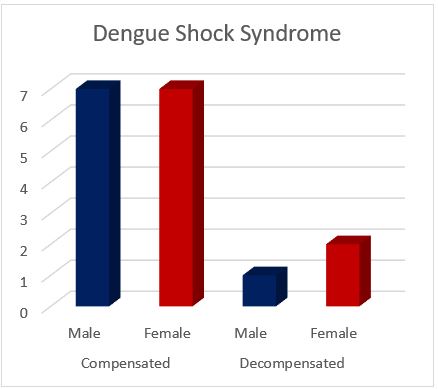
Figure 2 Gender wise versus type of Dengue Shock Syndrome Distribution.
Ascites (Figure-3c) was found in 19% (n=45) of the patients with mild ascites in 25 patients, moderate in 16 while sever in 4 patients. Graph in Figure 3-d shows the percentage wise abdominal ultrasound findings. Chest ultrasound showed pleural effusion in 28 (n=11.8%) patients with 21 having mild effusion while seven having moderate pleural effusion.
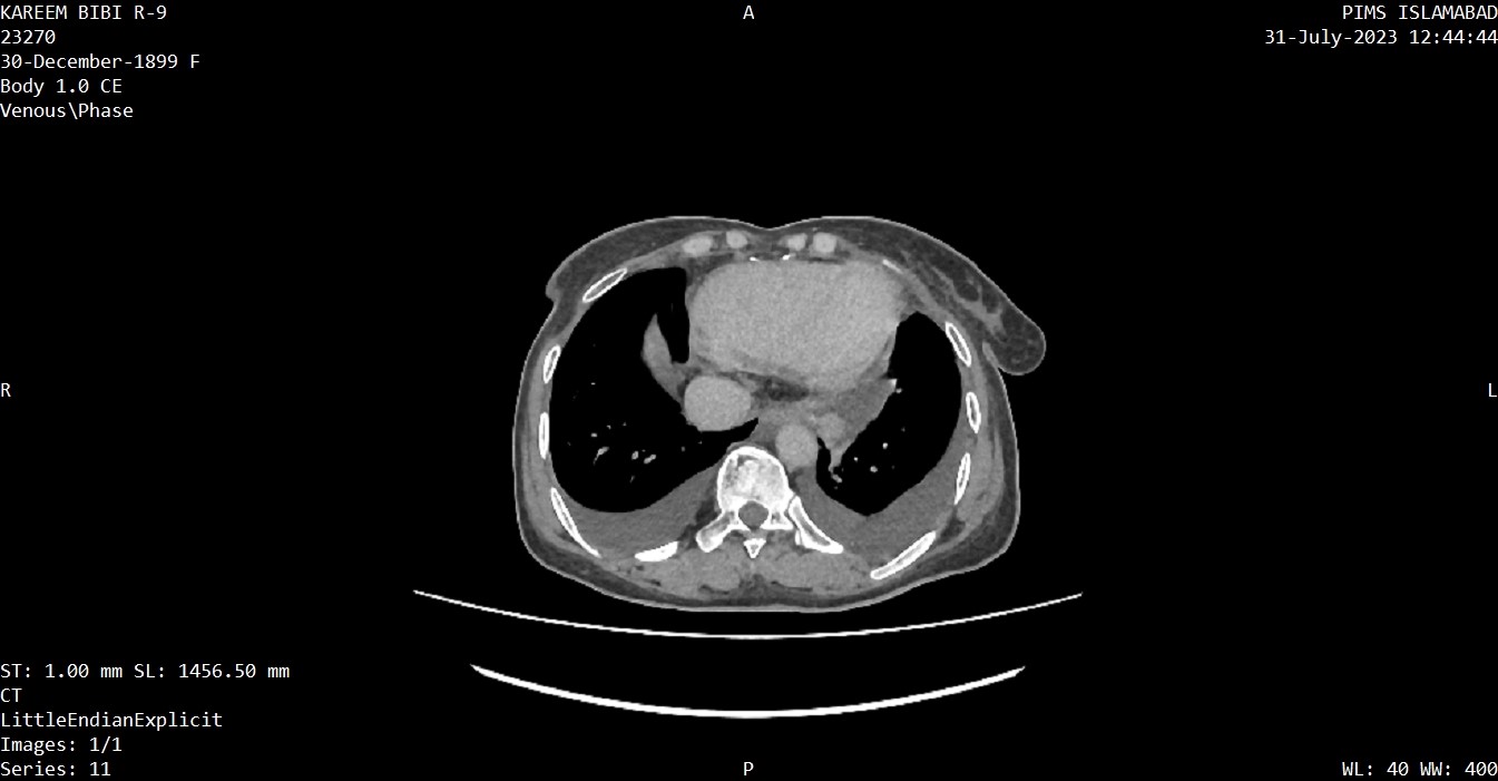
Figure 3: showing bilateral pleural effusion.
CT brain was performed in 3 patients, out of which two had intracerebral bleed. No other finding was observed on CT Brain.
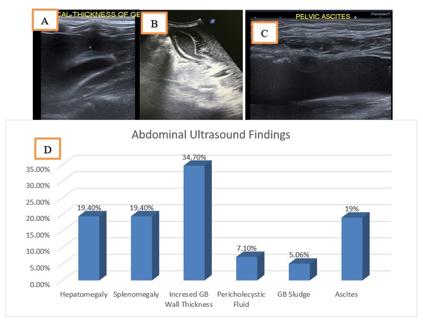
Figure 4: (a) shows focal GB wall thickening, (b) showing pericholecystic fluid, (c) pelvic ascites and (d) shows Percentage wise Abdominal Ultrasound Findings
The aim of this retrospective study was to investigate the spectrum of imaging findings in patients with Dengue Hemorrhagic Fever (DHF). Our findings provide valuable insights into the imaging manifestations and associated clinical parameters in a cohort of 237 patients diagnosed with DHF. The mean age of the patients in our study was 34.50 ± 16.4, with a male preponderance. These demographics are consistent with previous studies reporting a higher prevalence of DHF in adult males in Pakistan 1,2. The mean platelet count at the time of imaging was 98645.41 ± 88110.05, indicating thrombocytopenia, a characteristic feature of DHF. We classified the severity of DHF based on the World Health Organization (WHO) criteria, with Grade I DHF observed in 69.2% of the patients, followed by Grade II in 23.6% and Grade III in 7.1% of the patients. Additionally, 7.1% of the patients experienced shock, with three cases of decompensated shock and 14 cases of compensated shock. These findings underscore the importance of timely recognition and management of shock in DHF patients. Abdominal ultrasound revealed several notable findings. Hepatomegaly was observed in 19.4% of the patients, while splenomegaly was present in 19.6% of the cases. The most common finding was a thick-walled gall bladder, detected in 34.7% of the patients. Similar ultrasound findings have been reported in a previous study in Pakistan (9). The presence of pericholecystic fluid and gall bladder sludge was observed in a smaller proportion of patients. Ascites, a common feature in severe DHF, was identified in 19% of the cases, with varying degrees of severity. Most of the reported studies have also presented these conditions as most common ultrasound findings i.e., ascites and thick-walled gall bladder, however, the frequency varies10 Chest ultrasound findings showed pleural effusion in 11.8% of the patients, with most cases classified as mild effusion. A subset of patients (n=19) underwent HRCT scans, which revealed additional pulmonary manifestations. Pleural effusion was observed in 17 patients, with varying degrees of severity. Pleural effusion is a known complication of severe dengue and can contribute to respiratory distress in affected individuals7. Ground glass opacities and consolidations were present in 7 patients, indicating lung parenchymal involvement. Alveolar shadowing and signs of non-cardiogenic oedema were also identified in a smaller number of patients. These findings highlight the potential role of HRCT in detecting pulmonary complications in DHF cases. In our study, CT brain scans were performed in 3 patients with neurological manifestations. Intracerebral bleed was detected in two cases, emphasizing the occurrence of haemorrhagic manifestations in severe dengue cases. No other significant findings were observed on CT brain scans in our cohort. These findings contribute to the understanding of the imaging spectrum in patients with DHF, providing important clinical insights. The imaging modalities employed in this study, including abdominal ultrasound, chest ultrasound, HRCT, and CT brain scans, play a valuable role in the evaluation and management of DHF patients, aiding in the detection of complications and guiding appropriate interventions. It is important to acknowledge the limitations of our study. Firstly, the retrospective nature of the study introduces inherent biases and limitations in data collection. Additionally, the sample size was relatively small, which may limit the generalizability of our findings. Future prospective studies with larger sample sizes are warranted to validate and expand upon our results.
In conclusion, our retrospective study provides valuable insights into the spectrum of imaging findings in patients with Dengue Hemorrhagic Fever (DHF). We observed various imaging manifestations in abdominal and chest ultrasound, HRCT scans, and CT brain scans, which contribute to a better understanding of the disease's clinical presentation. Abdominal ultrasound revealed hepatomegaly, splenomegaly, and gall bladder abnormalities, while chest ultrasound and HRCT scans identified pleural effusion and pulmonary complications. CT brain scans detected intracerebral bleeding in patients with neurological manifestations. The utilization of imaging modalities assists in the evaluation and management of DHF cases, enabling timely interventions and appropriate care. Future prospective studies with larger sample sizes are warranted to validate our findings and enhance our understanding of the imaging features in DHF.
- Rehman AU, Anwar F, Tayyab M, Haq I, Haq M, Ahmed A, et al. Incidence of Dengue fever, serotypes, clinical features, and laboratory markers: a case study of 2019 outbreak at district Shangla, KP, Pakistan. Afr Health Sci. 2022;22(1):521–31. https://doi.org/10.4314/ahs.v22i1.61
- Anwar F, Tayyab M, Salman M, Abdullah, Din M, Khan J, et al. Dengue outbreak 2018 in district Shangla KPK; clinical features and laboratory markers of dengue virus infection. https://doi.org/102217/fvl-2019-0130 [Internet]. 2020 Oct 27 [cited 2023 Jul 3];15(10):693–9. http://dx.doi.org/10.2217/fvl-2019-0130
- Khan J, Ghaffar A, Khan SA. The changing epidemiological pattern of Dengue in Swat, Khyber Pakhtunkhwa. PLoS One [Internet]. 2018 Apr 1 [cited 2023 Jul 3];13(4):e0195706. https://doi.org/10.1371/journal.pone.0195706
- Rehman AU, Anwar F, Tayyab M, Haq I, Haq M, Ahmed A, et al. Incidence of Dengue fever, serotypes, clinical features, and laboratory markers: a case study of 2019 outbreak at district Shangla, KP, Pakistan. Afr Health Sci [Internet]. 2022 Apr 29 [cited 2023 Jul 3];22(1):521–31. https://doi.org/10.4314/ahs.v22i1.61
- Ali A, Fatima Z, Wahid B, Rafique S, Idrees M. Cosmopolitan A1 lineage of dengue virus serotype 2 is circulating in Pakistan: A study from 2017 dengue viral outbreak. J Med Virol [Internet]. 2019 Nov 1 [cited 2023 Jul 3];91(11):1909–17. https://doi.org/10.1002/jmv.25537
- Kumar A, Sahay N, Singh S, Singh K, Tiwari S. Dengue Hemorrhagic Fever with Pulmonary Complication in a Foreigner: A Case Report with a Brief Review of the Literature. Journal of Research & Innovation in Anesthesia [Internet]. 2023 May 22;8(1):23–5. https://doi.org/10.5005/jp-journals-10049-2022
- Kaagaard MD, Matos LO, Evangelista MVP, Wegener A, Holm AE, Vestergaard LS, et al. Frequency of pleural effusion in dengue patients by severity, age and imaging modality: a systematic review and meta-analysis. BMC Infect Dis [Internet]. 2023 Dec 1 [cited 2023 Jul 3];23(1):327. https://doi.org/10.1186/s12879-023-08311-y
- Awal MdR, khandakar MdIH, Chowdhury A, Islam MS. A case report of critically ill COVID-19 ICU patient recovery from acute respiratory distress syndrome, acute heart failure, kidney disorder, and Churg-Strauss syndrome. Authorea Preprints [Internet]. 2022 Dec 7 [cited 2023 Jul 3]; https://doi.org/10.22541/au.167041263.36399991/v1
- Bashir Z, Kamran AU, Saeed H, Nazir R, Patras S, Mehmood K, et al. Ultrasound Findings in Dengue Fever: A Single Center Study. Pakistan Journal of Medical and Health Sciences. 2022 Apr 26;16(4):756–8. https://doi.org/10.53350/pjmhs22164756
- Dewan N, Zuluaga D, Osorio L, Krienke ME, Bakker C, Kirsch J. Ultrasound in Dengue: A Scoping Review. Am J Trop Med Hyg [Internet]. 2021 Jan 18 [cited 2023 Jul 3];104(3):826. https://doi.org/10.4269/ajtmh.20-0103
- Wang WH, Urbina AN, Chang MR, Assavalapsakul W, Lu PL, Chen YH, et al. Dengue hemorrhagic fever – A systemic literature review of current perspectives on pathogenesis, prevention and control. Journal of Microbiology, Immunology and Infection. 2020 Dec 1;53(6):963–78. https://doi.org/10.1016/j.jmii.2020.03.007

An Official Publication of
Islamabad Medical & Dental College
Volume 13 Issue 2
Address of Correspondence
Ramish Riaz
Email:
ramish.riaz@riphah.edu.pk
Authors’ Contribution: 1,2Conception; Literature research; manuscript design and drafting; 3,4 Critical analysis and manuscript review; 5,6Data analysis; Manuscript Editing.
Cite this article. Akhtar S, Khatoon S, Raza M, Majeed AI, Shah SZ, Qazi A, Riaz R. Radiological Evaluation of Various Complications of Dengue Hemorrhagic Fever. J Islamabad Med Dental Coll. 2024; 13(2): 277-282.
DOI: https://doi.org/10.35787/jimdc.v13i2.1052