Gallbladder Perforation as a Complication of Enteric Fever: An Intriguing Case of Acute Abdomen
Aleena Tahir1, Mehwish Tabassum1, Wajeeha Anum1, Saba Khilji1, Manal Niazi1, Mashaal Shabrani2
1Department of Radiology, Dr. Akbar Niazi Teaching Hospital Islamabad, 2Sindh Institute of Urology and Transplantation, Karachi
If not identified and treated right once, gallbladder perforation—an uncommon but dangerous consequence of acute cholecystitis—can result in significant morbidity and mortality. We describe the example of a female patient, 19 years of age, who had a history of constipation. The patient was diagnosed with typhoid fever and underwent imaging studies that revealed gallbladder wall perforation along its posteroinferior aspect with adjacent contained right subhepatic collection. The patient underwent diagnostic laparoscopy and subtotal cholecystectomy. This instance emphasizes how crucial it is to rule out gallbladder perforation while diagnosing acute abdomen in typhoid fever patients. Prompt surgical intervention and early diagnosis can avert major problems and enhance results.
If not identified and treated right once, gallbladder perforation—an uncommon but dangerous consequence of acute cholecystitis - can result in significant morbidity and mortality.1 It is more common in elderly patients and those with comorbidities but can also occur in young patients with no underlying medical conditions.2
In underdeveloped nations, Salmonella typhi-induced enteric fever is a frequent cause of severe febrile sickness and can result in complications such as gallbladder rupture.3
We present a 19-year-old female patient in this case report who had a 10-day history of fever and abdomen pain. Positive results for Salmonella typhi IgG and IgM tests led to the diagnosis of enteric fever, and imaging examinations showed a gallbladder wall rupture with a nearby subhepatic collection.
The patient underwent diagnostic laparoscopy followed by exploration laparotomy and subtotal cholecystectomy and was managed postoperatively with antibiotics and supportive care. This case highlights the significance of surgical intervention in the treatment of gallbladder perforation as well as the role that imaging tests like contrast-enhanced CT and ultrasonography play in the identification of this problem.
Case Representation: A 19-year-old female patient arrived with a 10-day history of fever and abdomen
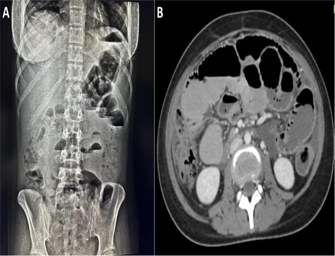
Figure 1: A) Dilated tiny gut loops and various air fluid levels associated with a minor bowel blockage; B) Axial CECT abdomen demonstrating multiple dilated gut loops with air fluid levels
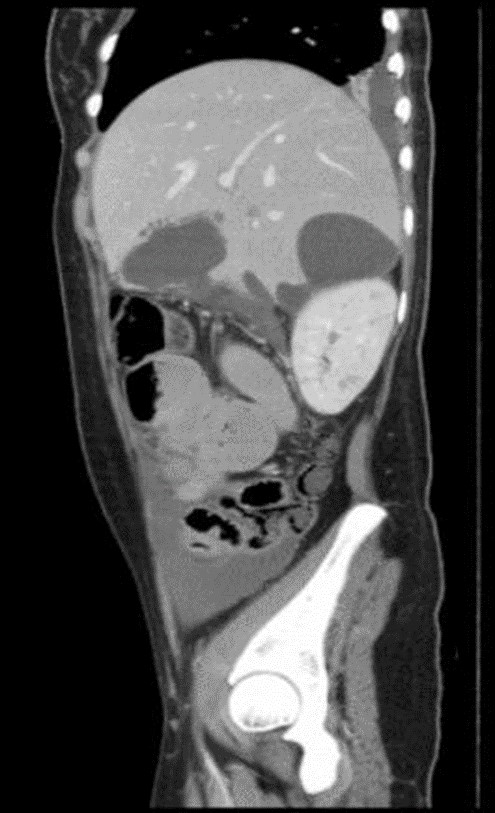
Figure 2: Coronal CECT abdomen showing gall bladder perforation with multiple contained intra-hepatic collections. Small focal defect visualized in posterior wall through which bile contents seen tracking posteroinferiorly into the right sub hepatic space forming collection between liver and kidney. Abdomino-pelvic free fluid collection also appreciated.
The 19-year-old female patient in the case study had a history of constipation, vomiting, fever, nausea, and abdomen pain. Based on the patient's clinical presentation and the results of the laboratory tests, enteric fever was the diagnosis made.
However, her condition worsened, and she developed signs of peritonitis. An emergency laparotomy was performed, which revealed a perforated gallbladder. the patient underwent subtotal cholecystectomy and made an uneventful recovery. Salmonella typhi or Salmonella paratyphi, which is widespread in many developing countries,
Table I: The Gallbladder Perforation Classification System by Niemeier: Categorizing Severity and Clinical Implications |
|||
Type |
Description |
Severity |
Clinical Features |
Type I |
Acute-free perforation causes peritonitis due to bile spillage into the peritoneal cavity. High mortality. |
Most severe |
Severe peritonitis, high mortality |
|
|
|
|
Type II |
Subacute or localized perforation causes abscesses in the vicinity of the gallbladder. |
Moderate severity |
Localized abscesses, |
Type III |
Moderate, chronic symptoms, and a persistent fistula forming between the gallbladder and nearby organs. |
Least severe |
Cholecystoenteric or cholecystocolic fistula |
can cause enteric fever, a systemic illness.4 It might cause fatal consequences such as intestinal perforation, sepsis, and diarrhoea in addition to the usual symptoms of fever, abdominal discomfort, and diarrhoea.5 Enteric fever can have rare but potentially fatal complications, including gallbladder perforation, which can be hard to identify because of its vague symptoms6. Gallbladder perforations were categorized by Niemeier into three kinds (Table I). Imaging investigations, laboratory results, and clinical presentation is used to diagnose enteric
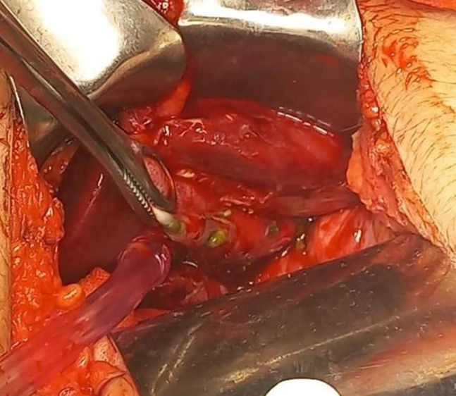
Figure 3: Multiple perforations were noted within the gall bladder. Surgical drain placed seen in situ.
The preferred surgical procedure for treating gallbladder perforation in enteric fever is cholecystectomy. In cases of perforation, subtotal cholecystectomy may be performed to avoid injury to the common bile duct. Antibiotics are also given to treat the underlying infection.10 A rare but possibly fatal consequence of enteric fever is gall bladder perforation. Prompt surgical intervention and early diagnosis can avert potentially fatal consequences.
If left untreated, gallbladder perforation can lead to significant morbidity and mortality. In underdeveloped nations, Salmonella typhi-induced enteric fever is a common cause of severe febrile sickness and complications like gallbladder rupture. Managing gallbladder perforation as an enteric fever complication necessitates early diagnosis and swift surgical intervention. Diagnostic imaging, including contrast enhanced CT, abdominal radiography, and ultrasonography, aids in diagnosing gallbladder perforation. This case study underscores the importance of considering gallbladder perforation as a potential enteric fever consequence, emphasizing the urgency of identification and treatment to prevent severe morbidity and fatality.
- Warsinggih, Mudatsir, Arsyad A, Faruk M. Gallbladder perforation: A rare case report. Int J Surg Case Rep. 2023 Mar 1; 104:107927. https://doi.org/10.1016%2Fj.ijscr.2023.107927
- Sheoran SK, Sahai RN, Indora J, Biswal UC. Spontaneous Perforation of Gallbladder: Case Report. Gastroenterology Res. 2016;9(2–3):61–3. https://doi.org/10.14740%2Fgr702e
- Yemeli Piankeu AD, Fodouop SPC, Noubom M, Gomseu Djoumsie EB, Ful Kuh G, Gatsing D. Epidemiology and Performances of Typhidot Immunoassay and Widal Slide Agglutination in the Diagnosis of Typhoid Fever in Febrile Patients in Bafoussam City, Cameroon: A Cross-Sectional Comparative Study. Can J Infect Dis Med Microbiol. 2024 ;2024:6635067. https://doi.org/10.1155/2024/6635067.
- Antillón M, Warren JL, Crawford FW, Weinberger DM, Kürüm E, Pak GD, Marks F, Pitzer VE. The burden of typhoid fever in low- and middle-income countries: A meta-regression approach. PLoS Negl Trop Dis. 2017;11(2):e0005376. https://doi.org/10.1371/journal.pntd.0005376.
- Obasi AA, Igboanugo AA. Gall Bladder Complications Resulting from Typhoid Fever in Children: Challenges of Management and Lessons Learned. J West Afr Coll Surg. 2020;10(1):35-38. https://doi.org/10.4103/jwas.jwas_31_21.
- Malik MN, Mahmood T, Tameez Ud Din A, Aslam S, Imtiaz M. Gallbladder Perforation Secondary to Enteric Fever: An Interesting Case of Acute Abdomen. Cureus. 2019 Apr 22;11(4): e4516. https://doi.org/10.7759%2Fcureus.4516
- Case reports. J Family Med Prim Care. 2018;7(9):27. http://dx.doi.org/10.15761/HPC.1000146
- Bhandari TR, Khan SA, Jha JL, Sah JK. A rare case report of enteric fever causing gallbladder perforation. Int J Surg Case Rep. 2021 Nov 1; 88:106553. https://doi.org/10.1016%2Fj.ijscr.2021.106553
- Subhan M, Sadiq W. Case of Enteric Fever with Bicytopenia. Cureus. 2017 Dec 6. http://dx.doi.org/10.7759/cureus.1910
- Singh M, Kumar L, Singh R, Jain AK, Karande SK, Saradna A, et al. Gallbladder perforation: A rare complication of enteric fever. Int J Surg Case Rep. 2014 Jan 1;5(2):73–5. https://doi.org/10.1016%2Fj.ijscr.2013.12.004

An Official Publication of
Islamabad Medical & Dental College
Volume 13 Issue 2
Case Report
Address of Correspondence
Aleena Tahir
Email:
aleenatahir091095@gmail.com
Authors’ Contribution: 1,2Conception; Literature research; manuscript design and drafting; 3,4 Critical analysis and manuscript review; 5,6 Data analysis; Manuscript Editing.
Cite this article. Tahir A, Tabassum M, Anum W, Khilji S, Niazi M, Shabrani M. Gallbladder Perforation as a Complication of Enteric Fever: An Intriguing Case of Acute Abdomen.J Islamabad Med Dental Coll. 2024; 13(2): 386-389.
DOI: https://doi.org/10.35787/jimdc.v13i2.1086