Pulmonary Cyst: A Rare Extra-Renal Manifestation of Autosomal Dominant Polycystic Kidney Disease
Humaira Riaz1, Fizza Batool2, Hina Hanif Mughal2, Zainab Riaz3, Ania Javed4, Sharjeel Sarfaraz1
1Rawalpindi Medical University, Institute of Urology and Transplantation; 2Benazir Bhutto Hospital Rawalpindi; 3Islamabad Diagnostic Center, Islamabad; 4District Headquarter Hospital, Haripur
Autosomal dominant polycystic kidney disease (ADPKD) is characterized by bilateral multiple renal cysts of varying sizes leading to end stage renal failure over the subsequent years. Though there is a wealth of information regarding the extrarenal visceral linkages of ADPKD, very few studies have described constellate pulmonary findings within the spectrum of extrarenal manifestations. This case report features a 55-year-old woman who had intermittent flank pain for about two years until an ultrasound revealed she had ADPKD. HRCT chest revealed pulmonary cysts in bilaterally lung fields. Patient had no pre-existing pulmonary disease or co-existing risk factors; HRCT findings were considered to be the spectrum of ADPKD associations. The report underscores the need for comprehensive systemic diagnostic evaluation in patients for ADPKD, as fatal complications like pneumothorax can be the first presentation of such patients, besides other systemic complaints.
Keywords: Extra-renal, hyperdense cyst, Pulmonary cyst, dialysis
About 500,000 people suffer from the systemic condition known as autosomal dominant polycystic kidney disease (ADPKD), which makes up 5–10% of the dialysis population in the US alone.1 Around half of those with ADPKD will have advanced end-stage renal disease by the time they are 60 years old. ADPKD often first appears in the third or fourth decade of life.2 ADPKD frequently presents with extra-renal symptoms, such as liver, pancreatic, central nervous system, and genitourinary tract cysts.3 Bronchiectasis has been the most prevalent pulmonary manifestation of ADPKD in the few published cases with synchronous lung disease; even fewer case reports describe concomitant pulmonary cysts.4 Here, we describe a rare instance of ADPKD's pulmonary manifestation—cystic lung disease.
A 55-year-old female patient arrived at our hospital's Department of Urology outpatient clinic with 2-year history of right flank pain. Initially it was on and off but had become constant for the last 2 months. Her past urological history was remarkable for recurrent urinary tract infections treated successfully with short courses of antibiotics.
When she presented it to our department her vitals were within normal limits except blood pressure which was 130/90 mmHg. General physical examination showed mild tenderness in both flank regions. Ultrasonography of abdomen demonstrated bilateral enlarged kidneys showing significant replacement of renal parenchyma by innumerable variable sized cysts, few of them showing foci of calcification. Most of the cysts were predominantly anechoic, however a few were hemorrhagic cysts. Two renal calculi were seen in right kidney, larger being present at renal pelvis. Two small calculi were also in left kidney. The rest of the abdominal viscera including liver and pancreas appeared unremarkable with no evidence of co-existing cysts. Non-contrast CT (NCCT) KUB showed bilateral moderately enlarged kidneys with multiple cysts, ranging in size from a few millimeters to multiple centimeters.
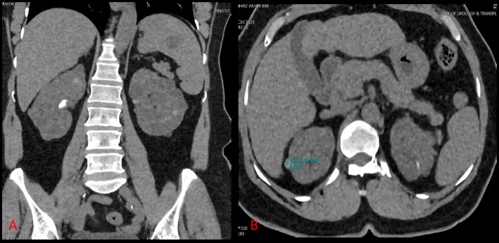
Figure 1A: Bilateral multiple variable sized renal cysts varying in density; most are near-water density, some are hyperdense (1B), few showing marginal calcification. Stone 7 mm (501 HU) evident at right renal collecting system. hypodense area in spleen likely splenic cyst.
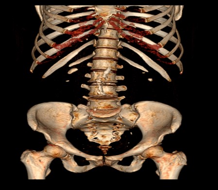
Figure 2: Bone window showing bilateral renal calculi. Atherosclerotic calcification of abdominal aorta and its branches also noted.
A few cysts were showing calcification in their walls as shown in figure 2. Two renal calculi were noted in right kidney measuring 7.1 mm (501 HU) and 4.5 mm (467 HU) in size at renal pelvis and lower poles respectively. In left kidney calculi measures 7 mm (572 HU) and 4.4 mm (403 HU) as shown on 3D bone reconstruction in Error! Reference source not found.. The patient had no prior diagnostic evaluation of kidneys nor any positive family history of ADPKD. Visualized basilar segments of bilateral lower lung zones showed a few thin-walled pulmonary cysts. HRCT chest was done which revealed multiple pulmonary cysts in bilateral lung fields (Error! Reference source not found.). There was no evidence of pleural effusion or bronchiectasis changes on either side. The patient denied any smoking history, inhaled drug use, or inhaled allergen exposure. Based on patient’s history and imaging findings diagnosis of pulmonary cysts as rare extra-renal manifestation of autosomal dominant polycystic kidney disease (ADPKD) was made.
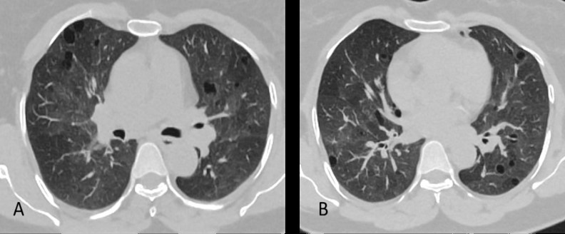
Figure 3: Axial plain HRCT chest images of the thorax at the level of mainstem bronchi (A), and in the lower lung zones (B) show bilateral pulmonary cysts with no lobar predominance, intervening normal lung parenchyma appreciated.
With an incidence of 1:500 to 1:1000, The most common kind of polycystic kidney disease is ADPKD. The formation of large bilateral renal cysts has been a well-characterized feature of ADPKD renal symptoms. In the third and fourth decades of life, it frequently shows up.5 By the time they are 60 years old, about 50% of individuals will have advanced to end stage renal disease due to the replacement of healthy renal parenchyma.
Mutations in the PKD1 or PKD2 genes, which encode for the polycystin 1 and 2 proteins, respectively, have been associated to the pathogenesis of ADPKD.6 Eighty to ninety percent of patients with ADPKD have been found to have PKD1 gene mutations; the remainder patients usually have PKD2 gene mutations, which usually result in a milder course of the disease.
Extra-renal associations of ADPKD include cerebral berry aneurysms, hepatic cysts, pancreatic cysts, seminal vesicle cysts and infertility.3 Bronchiectasis and pulmonary cyst development are the most frequently documented synchronous pulmonary diseases. These conditions are believed to be attributable to polycystin mutations that cause ciliary failure affecting the smooth muscle and airway epithelium.4
The most common sign of ADPKD in the lung is bronchiectasis, which has a reported frequency of 19–37 percent.4
Only a few case reports have described pulmonary associations of ADPKD which showed that the frequency of pulmonary cyst formation is significantly lower than that of bronchiectasis. All of the previously documented cases had lung cysts linked to underlying ADPKD since no other plausible diagnosis could be made. Because pulmonary cysts associated with ADPKD are uncommon, it has been suggested that the combination of renal and pulmonary cysts could be explained by concurrent tuberous sclerosis complex (TSC).7
Pulmonary langerhans cell histiocytosis, lymphangioleiomyomatosis (LAM), and lymphocytic interstitial pneumonia are among the differential considerations for cystic lung illness.8 Our patient did not fit in the traditional profile of LAM patients, who are typically women of childbearing age, and did not exhibit any recognizable symptoms of tuberous sclerosis, which makes LAM implausible.9 Since our patient did not smoke, lung langerhans cell histiocytosis and desquamative interstitial pneumonia were ruled out.10 Thus, it was determined that our patient's underlying ADPKD was the cause of her cystic lung condition.
Furthermore, the absence of any history of inhaled allergen exposure in our patient lowers the possibility of chronic hypersensitivity pneumonitis.11
This case report focuses on pulmonary cysts, an uncommon extra-renal symptom of autosomal dominant polycystic kidney disease (ADPKD). Despite the challenging diagnostic landscape, the clinical and radiological findings, along with the absence of identifiable alternative causes, led to the attribution of the pulmonary cysts to ADPKD in this patient. The report highlights the need for comprehensive evaluation and ongoing research to elucidate the relationship between ADPKD and pulmonary manifestations.
- Akbari M, West JD, Doerr N, Kipp KR, Marhamati N, Vuong S, et al. Restoration of atypical protein kinase C ζ function in autosomal dominant polycystic kidney disease ameliorates disease progression. Proc Natl Acad Sci U S A [Internet]. 2022 Jul 26 [cited 2024 Jan 14];119(30): e2121267119. https://doi.org/10.1073/pnas.2121267119
- Gallo-Bernal S, Kilcoyne A, Gee MS, Paul E. Cystic kidney disease in tuberous sclerosis complex: current knowledge and unresolved questions. Pediatric Nephrology [Internet]. 2023 Oct 1 [cited 2024 Jan 14];38(10):3253–64. https://doi.org/10.1007/s00467-022-05820-x
- Righini M, Mancini R, Busutti M, Buscaroli A. Autosomal Dominant Polycystic Kidney Disease: Extrarenal Involvement. 2024 Jan 4 [cited 2024 Jan 15]; https://doi.org/10.3390/ijms25052554
- Levy N, Hota P, Kumaran M. Coexisting cystic lung disease as a rare extra-renal manifestation of autosomal dominant polycystic kidney disease. Radiol Case Rep. 2018 Oct 1;13(5):1048–52. https://doi.org/10.1016/j.radcr.2018.04.013
- Rangan G, Wong A, Munt A, Sangadi I, Saravanabavan S, Zhang J. Autosomal Dominant Polycystic Kidney Disease. Evidence‐Based Nephrology [Internet]. 2022 Dec 2 [cited 2024 Jan 15];288–304. https://doi.org/10.1002/9781119105954.ch19
- Ong ACM, Harris PC. Molecular pathogenesis of ADPKD: The polycystin complex gets complex. Kidney Int. 2005 Apr 1;67(4):1234–47. https://doi.org/10.1111/j.1523-1755.2005.00201.x
- Nair N, Chakraborty R, Mahajan Z, Sharma A, Sethi SK, Raina R. Renal Manifestations of Tuberous Sclerosis Complex. J Kidney Cancer VHL [Internet]. 2020 Aug 27 [cited 2024 Jan 15];7(3):5. https://doi.org/10.15586/jkcvhl.2020.131
- Raoof S, Bondalapati P, Vydyula R, Ryu JH, Gupta N, Raoof S, et al. Cystic Lung Diseases: Algorithmic Approach. Chest. 2016 Oct 1;150(4):945–65. https://doi.org/10.1016/j.chest.2016.04.026
- Baskin HJ. The pathogenesis and imaging of the tuberous sclerosis complex. Pediatr Radiol [Internet]. 2008 Sep 15 [cited 2024 Jan 15];38(9):936–52. https://doi.org/10.1007/s00247-008-0832-y
- Vancheri C, Puglisi S. Pulmonary Langerhans Cell Histiocytosis and Smoking-Related Interstitial Lung Diseases. Orphan Lung Diseases [Internet]. 2015 [cited 2024 Jan 15];435–56. https://doi.org/10.1513/pats.200512-135tk
- Patel AM, Ryu JH, Reed CE. Hypersensitivity pneumonitis: Current concepts and future questions. Journal of Allergy and Clinical Immunology. 2001 Nov 1;108(5):661–70. https://doi.org/10.1067/mai.2001.119570

An Official Publication of
Islamabad Medical & Dental College
Vol 13 No (Suppl.)
Case Report
Address of Correspondence
Huma Riaz
Email:
drhumaira2801@gmail.com
Authors’ Contribution: All authors contributed equally to the conception, literature search, manuscript drafting, editing and review
Cite this article. Riaz H, Batool F, Mughal HH, Riaz Z, Javed A, Sarfraz S. Pulmonary Cyst: A Rare Extra-Renal Manifestation of Autosomal Dominant Polycystic Kidney Disease. J Islamabad Med Dental Coll. 2024; 13i(Suppl.): 574-577.
DOI: https://doi.org/10.35787/jimdc.
v13i(Suppl.)1101.