Emphysematous Spinous Osteomyelitis and Acute Pyelonephritis in Diabetic Patient
Mariam Shah, Maira Aslam, Khurram Khaliq Bhinder, Madiha Saeed Wahla, Aroosa Kanwal
Department of Radiology, Shifa International Hospital Islamabad.
Spontaneous emphysematous osteomyelitis with concurrent pyelonephritis is exceedingly rare and fatal entity that needs prompt diagnosis. Imaging plays a crucial role in its diagnosis. We present a case of 52 years old woman known case of diabetes and hypertension presenting to emergency department with high grade fever and right flank pain. CT scan abdomen and pelvis revealed intraosseous gas extending in epidural space and in retro peritoneum, suggesting emphysematous osteomyelitis, which was later confirmed by E coli in blood culture. Right kidney was also swollen and enlarged with surrounding perinephric stranding. This is a rare case and highlights the importance of radiological imaging in Emergency settings particularly in patients with comorbid. Being familiar with this rare entity thus can help in early diagnosis and initiation of treatment.
Keywords: Emphysematous osteomyelitis, Pyelonephritis, Diabetes, Computed tomography.
Emphysematous osteomyelitis is infection of bone caused by gas forming organism with a very high mortality rate of 32%. Only 10 cases are described so far involving the spine. The presence of intra-osseous air, when aligned with appropriate clinical scenario, provide significant evidence for diagnosis. However, it is important to exclude other causes of intra-osseous air such as trauma, degenerative changes etc.1
The co-existence of emphysematous osteomyelitis and acute pyelonephritis is life threatening occurring exclusively in patients with known comorbid like diabetes and is even rarer with 3 cases seen previously.2
The co-existence of both diseases is seen in diabetics as in our case and if not recognized and treated early can lead to sepsis and eventually death.3
To our best knowledge, no case has been described from Pakistan.
Here, we report a case of 52 years old with poorly controlled diabetes myelitis, who presented with emphysematous osteomyelitis and concurrent right sided acute pyelonephritis with blood culture positive for E-coli.
A 52 years old female known diabetic and hypertensive presented to ER with highly grade fever from 2 weeks. She also has burning micturition, urinary urgency, frequency and right flank plain and was suspected as pyelonephritis. On arrival patient was hypotensive and had tachycardia. On examination she had right flank tenderness and moderate dehydration. Ct scan abdomen and pelvis was performed in ER and revealed extremely swollen right kidney with perinephric stranding. (Figure 1). No air with renal parenchyma or collecting system was seen and spine showed extensive emphysema in spinal canal along with intraosseous air in L2 and L3 vertebral bodies without any evidence of vertebral fracture.
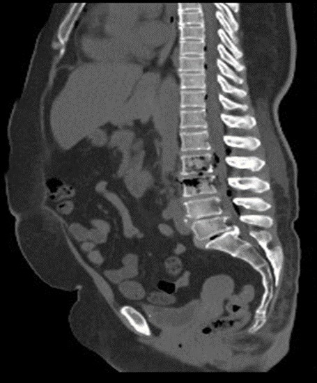
Figure 1. CT abdomen sagittal view showing intra-osseous air in L2 and L3 vertebral bodies extending into intra-vertebral and epidural space.

Figure 2. 2CT scan abdomen showing significantly swollen right kidney with mild peri-nephric stranding. Intra-osseous air extending into retroperitonuem is also seen.
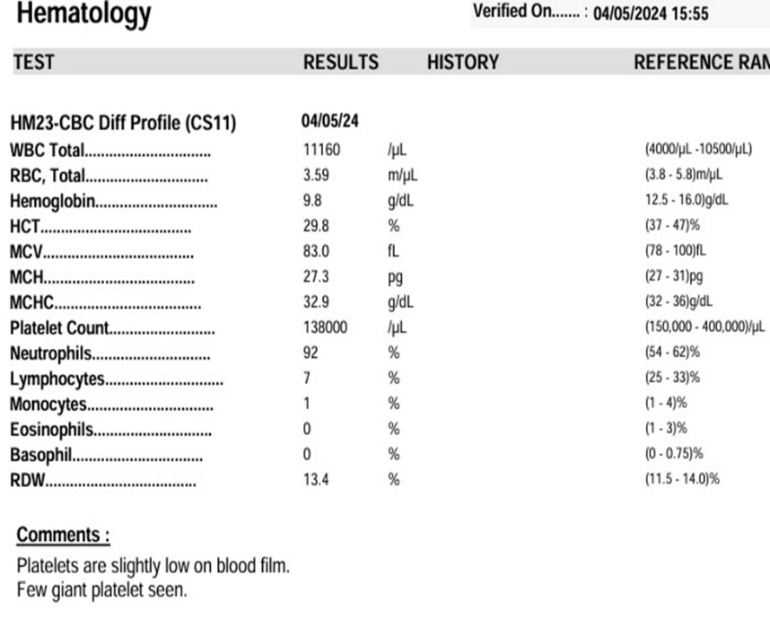
Figure 3. Hematology report
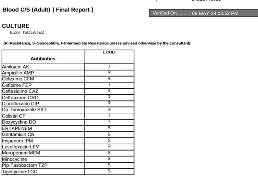
Figure 4. Blood culture report
Lab workup revealed anemia (red blood cells= 3.59 x10^5), thrombocytopenia (platelet count=13 x 10^3), leukocytosis (white blood cells=11160), high C Reactive protein (205mg/dl) and hyperglycemia (glucose=270mg/dl) (Figure 3). Urine dipstick showed presence of glucose +++, blood ++++ and leukocytes ++. Blood culture of patient was positive for E coli in Figure 4.
Emphysematous osteomyelitis of spine is extremely rare with very few cases reported in literature so far. Having said that identification of emphysematous osteomyelitis is crucial since it carries high morbidity and mortality. Gas collection in axial skeleton without any preceding history of trauma or surgery in an appropriate clinical setting warrants to consider infection as a potential culprit. There is no sex predilection with 51 years being the median presenting age 4. Various immune deficient states have been considered as important contributing risk factors.
While diagnosis of osteomyelitis can be made on clinical signs and symptoms, radiological examination is almost always required for definite diagnosis. CT remains the gold standard for identification of intraosseous gas and describing the extent of involvement. The pattern of gas involvement also helps in isolating aggressive/ infectious causes from more commonly encountered benign causes. Intraosseous gas with linear pattern without any soft tissue abnormality is more likely because of benign causes whereas bubbly and irregular distribution is more convincing of emphysematous osteomyelitis.5 Another described pattern suggestive of emphysematous osteomyelitis is multiple variable sized gas foci in intramedullary cavity labelled as ‘pumice stone pattern.6 MRI can also be considered with advantage of detecting bone marrow and spinal cord involvement. However, it is restricted by long scan times on unstable patients and limited availability.
Anaerobes are mostly described as a causative organism just like in other gas forming infections, whereas the possibility of microbial infection also remains.1
When treating spinal osteomyelitis, it is crucial to identify the microbe and give targeted therapy. However, most reported cases have started with empirical antibiotic therapy.7
Diabetes is a common cause of both emphysematous and non-emphysematous pyelonephritis with E-coli being the most common responsible organism. The severity of urinary tract infection is also increased in diabetics and pyelonephritis is also often bilateral.8
Diagnosis of urinary tract infection is based on clinical features and lab work-up with imaging reserved for atypical and complicated cases. Although ultrasound can be used for diagnosing the condition CT proves to be more valuable as it also provides details about extrarenal extent of disease which aids in surgical planning.9
In our case report, we describe a patient with medical history of diabetes myelitis, presented to ER with high grade fever and Ct abdomen and pelvis revealing pyelonephritis and emphysematous osteomyelitis. E. coli was isolated from the blood cultures of our patient considering there was ongoing pyelonephritis which was most likely responsible of causing emphysematous osteomyelitis. This case highlights importance of CT in diagnosing these conditions. The case gives us some insight in pathophysiology of these condition with hyperglycemia being a causal factor
This case report underscores the role of radiology in timely diagnosis of unusual yet highly lethal complication of pyelonephritis. Clinical information can often be sparse and imaging plays a key role. It reinforces the facts that spinal osteomyelitis requires careful evaluation of retroperitoneal structures and can also present in emphysematous form. Highlighting this potentially fatal complicacy will prove helpful for the future similar clinical setting, thus decreasing the overall morbidity and mortality.
- Tatakis FP, Kyriazis I, Panagiotopoulou IE, Kalafatis E, Mantzikopoulos G, Polyzos K, Kachrimanidis I, Vogiatzakis AM, Rellou S, Manta E, Tzaki M. Simultaneous diagnosis of emphysematous osteomyelitis and emphysematous pyelonephritis in a diabetic patient. The American Journal of Case Reports. 2019; 20:1793. https://doi.org/ 10.4103/jpgm.jpgm_234_22
- Neelakandan S, Viswanathan S, Selvaraj J, Pillai V, Sharma D, Chakkalakkoombil SV. Concurrent presentation of emphysematous pyelonephritis, emphysematous osteomyelitis, and psoas abscesses. Cureus. 2021 Jun 24;13(6) https://doi.org/ 10.7759/cureus.15908
- Sninate S, Oubaddi T, Allioui S, Jroundi L, Laamrani FZ. Emphysematous pyelonephritis and emphysematous osteomyelitis: a case report. Galician Medical Journal. 2022 Mar 1;29(1):E202217 https://doi.org/10.21802/gmj.2022.1.7
- Ono R, Uehara K, Kitagawal I. Emphysematous osteomyelitis of the spine: a case report and literature review. Internal Medicine. 2018 Jul 15;57(14):2081-7. https://doi.org/10.2169/internalmedicine.0219-17
- Feng SW, Chang MC, Wu HT, Yu JK, Wang ST, Liu CL. Are intravertebral vacuum phenomena benign lesions? European Spine Journal. 2011 Aug; 20:1341-8. https://doi.org/10.1007/s00586-011-1789-3
- Small JE, Chea P, Shah N, Small KM. Diagnostic features of emphysematous osteomyelitis. Current Problems in Diagnostic Radiology. 2022 Jul 1;51(4):666-72. https://doi.org/10.1067/j.cpradiol.2018.05.008
- Lee D, Mohile N, Rakoczy K, Gjolaj JP. Emphysematous osteomyelitis of the spine: a case report and case-based review of the literature. Journal of Spine Surgery. 2022 Sep;8(3):362. https://doi.org/10.21037/jss-22-6
- Kumar S, Ramachandran R, Mete U, Mittal T, Dutta P, Kumar V, Rathi M, Jha V, Gupta KL, Sakhuja V, Kohli HS. Acute pyelonephritis in diabetes mellitus: Single center experience. Indian journal of nephrology. 2014 Nov 1;24(6):367-71. https://doi.org/10.4103/0971-4065.135347
- Craig WD, Wagner BJ, Travis MD. Pyelonephritis: radiologic-pathologic review. Radiographics. 2008 Jan;28(1):255-76. https://doi.org/10.1148/rg.281075171

An Official Publication of
Islamabad Medical & Dental College
Vol 13 No (Suppl.)
Case Report
Address of Correspondence
Khurram Khaliq Binder
Email:
kkbhinder@yahoo.com
Authors’ Contribution: All authors contributed equally to the conception, literature search, manuscript drafting, editing and review
Cite this article. Shah M, Aslam M, Binder KK, Wahla MS, Kanwal A. Emphysematous Spinous Osteomyelitis and Acute Pyelonephritis in Diabetic Patient. J Islamabad Med Dental Coll. 2024; 13i(Suppl.): 571-581. DOI: https://doi.org/10.35787/jimdc.
v13i(Suppl.)1226.