Sudden Maternal Death from Suspected Amniotic Fluid Embolism and a Dead Baby Delivered with Natal Teeth
Faiza Kamran Ali 1, Feriha Fatima Khidri 2, Kamran Ali Shahani 3, Rafia Shah 4
1 Postgraduate trainee, Department of Gynaecology and Obstetrics, Isra University Hospital, Hyderabad
2 Lecturer, Biochemistry Department, Liaquat University of Medical and Health Sciences, Jamshoro
3 Head, Department of Pediatrics, Khairpur Medical College
4Lecturer, Prosthetic dentistry, Liaquat University of Medical and Health Sciences, Jamshoro
Amniotic fluid embolism (AFE) is a rare presentation in obstetric emergencies that carries great risk for the life of both mother and fetus. It is usually characterized by sudden cardiovascular collapse, respiratory distress and disseminated intravascular coagulation. Here we present a case of sudden death of a pregnant woman due to suspected AFE. We also present a rare finding of natal teeth in her deceased baby, which along with reported AFE in the mother, is an unlikely event in the medical literature.
Key words: Amniotic fluid embolism, natal teeth, pregnancy
Amniotic fluid embolism (AFE) is a rare and unpredictable obstetric emergency, and an important cause of maternal mortality in the developed countries. It is characterised by a triad of sudden cardiovascular collapse, respiratory distress and disseminated intravascular coagulation.1 It is clinically diagnosed early during labour with ruptured membranes and occurs consequently due to the entry of amniotic fluid, fetal skin cells, hair or debris into the maternal circulation. It has a reported incidence of 1: 15,200 to 1: 53, 800 among pregnancies.2 The presence of tooth/teeth in a child at birth is an uncommon condition of the oral cavity and is referred to as ‘natal teeth’.3 The lower primary central incisors are most commonly erupted natal teeth, however eruption of multiple natal teeth (> 2) is an extremely rare occurrence.4
A 35-year-old gravida 6 para 5 was received in Emergency of Gynaecology and Obstetrics Department, Isra University Hospital Hyderabad, with a history of sudden death. The attendants gave history that the patient experienced sudden uterine contractions and rupture of membranes about six hours ago. On her way to the hospital, she suddenly developed shortness of breath with a cough and collapsed in the vehicle within 5-10 minutes. She had completed her 41 weeks of gestation and was previously healthy. All her previous pregnancies were un-eventful and normal vaginal deliveries. She did not have a history of diabetes mellitus, chronic hypertension, preeclampsia/eclampsia, bleeding disorder, gestational hypertension, antepartum haemorrhage, fever or trauma. Her death was confirmed on arrival at the hospital, however, fetal heart sounds were audible on fetoscope. Immediate peri-mortem emergency Caesarean section (C-section) was performed. After giving incision in the uterus, surgeons observed abnormally excessive uterine bleeding and suspected defective coagulation. The likely clinical diagnosis was amniotic fluid embolism, a rare pregnancy complication causing sudden cardiac arrest to multiple organ damage with or without coagulopathy. A dead male baby weighing 3.2 kg was delivered. Placenta appeared normal on gross examination. General physical and systemic examination of the neonate revealed no abnormality. On oral examination, lips, tongue, palate and the floor of the mouth were normal in appearance. However, the baby had 4 natal teeth (2 maxillary and 2 mandibular), which is an extremely rare finding. Teeth were whitish opaque in colour, mature and immobile. The size of the crown and gingival appearance were normal. The face of the baby was symmetrical and no morphological abnormality was seen (Figure 1). The cause of the AFE could not be determined due to the refusal of post-mortem by the patient’s family.
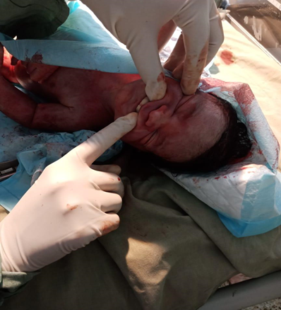
Figure 1: A new-born male baby with natal teeth in anterior maxillary area.
*(Photograph was taken after permission from the attendants)
The presented case describes the sudden death of a woman in labour with no identified known risk factors. The AFE diagnosis was made according to the criteria presented by Fitzpatrick et al.1. Lewis 5 reported that, 11 out of the 17 parturient women with experience of AFE presented with prodromal symptoms such as shortness of breath, pain in chest, agitation, pin pricking sensations, nausea and vomiting. The time period between prodromal symptoms to collapse ranged from minutes to four hours. In the present case report, the parturient woman experienced dyspnea accompanied with a cough; however, it lasted only for a few minutes followed by collapse with no other specific symptoms. AFE was diagnosed on the basis of sudden cardiac arrest, dyspnea, severe clinical bleeding observed during peri-mortem emergency C-section, and occurrence of the event during labour with no explanation for the clinical findings. Unfortunately, no pharmacological or other therapies presently have been able to prevent or treat AFE. It is managed with multidisciplinary approach using supportive treatment and managing shock simultaneously. 6
Table I: Diagnostic criteria for AFE |
In the absence of any other identified cause: |
OR |
According to previous studies, the mean age of patients presenting with SCC of renal pelvic area is around 56 years and usually there is no predilection for laterality or sex.6 In this case, the patient was a young male, aged 35 years, and tumor was present in the left kidney. This tumor usually arises from the urinary bladder, sometimes from the male urethra but very rarely from the renal pelvis. In patients presenting with this tumor, features of paraneoplastic syndromes have also been observed such as leukocytosis, hypercalcemia and thrombocytosis. Previously 84% of renal SCC presented as locally advanced or metastatic tumor.11 Various etiological factors have been associated with the pathogenesis of SSC of upper urinary tract but the strongest association has been noted with renal calculi. In the previously reported cases, up to 100% of renal SCC cases had nephrolithiasis.6 In this case, multiple renal calculi were also present7.
The occurrence of natal teeth was regarded as a bad omen in Chinese history, however, they were considered as a sign of luck in England, France and Italy. The mechanism behind natal teeth, although is not well defined, however, certain factors, such as superficial site of the germ, infections, malnutrition, febrile incidents, hormones, hereditary or unusual osteoblastic activity may confer risk. Its management to either extract or to retain, is usually decided by the dentition after assessing mobility and degree of maturity. 7
This case report diagnosed a case of amniotic fluid embolism on the basis of clinical criteria, which if undiagnosed poses a serious threat for both mother and baby. It also highlighted the unusual finding of multiple natal teeth in a new born baby. In the present case, it was an unfortunate event to receive a mother after death occurrence followed by delivery of a dead baby, therefore further management was not possible; though it provides awareness to clinicians about two rare events in the same case.
- Fitzpatrick KE, Tuffnell D, Kurinczuk JJ, Knight M. Incidence, risk factors, management and outcomes of amniotic‐fluid embolism: a population‐based cohort and nested case–control study. BJOG: An International Journal of Obstetrics & Gynaecology. 2016;123(1):100-9.
- Rezai S, Hughes AC, Larsen TB, Fuller PN, Henderson CE. Atypical Amniotic Fluid Embolism Managed with a Novel Therapeutic Regimen. Case Rep Obstet Gynecol 2017; ID 8458375
- Malki GA, Al-Badawi EA, Dahlan MA. Natal Teeth: A Case Report and Reappraisal. Case Rep Dent. 2015:21(40)4.
- Leung AK, Robson WLM. Natal teeth: a review. J Natl Med Assoc. 2006;98(2):226.
- Lewis G. The confidential enquiry into maternal and child health (CEMACH) The seventh report on confidential enquiries into maternal deaths in the United Kingdom. London: CEMACH. 2007.
- Moore J, Baldisseri MR. Amniotic fluid embolism. Crit Care Med 2005;33(10):S279-S85.
- Cunha RF, Boer FAC, Torriani DD, Frossard WTG. Natal and neonatal teeth: review of the literature. Pediatr Dent. 2001;23(2):158-62.
- Hebling J, Zuanon A, Vianna D. Dente Natal—a case of natal teeth. Odontol Clin. 1997;7:37-40.