Histomorphometric Alterations in Hepatic Tissue from Malathion-Induced Toxicity: An Experimental Animal Study
Shazia Parveen Channar1, Nasreen Qazi2, Sajjad Ali Almani3, Sehar Gul Memon4, Mansoor Mukhtar Qazi5, Rida Qureshi5
1Senior Lecturer, Department of Pharmacology, Isra University, Hyderabad Pakistan
2 Professor and Chairperson, Department of Pharmacology & Therapeutics, LUMHS, Jamshoro Pakistan
3 Assistant Professor, Department of Anatomy, Dow Medical University Karachi Pakistan
4 Lecturer, Department of Physiology, Ghad College, Riyadh, Saudi Arabia
5 Lecturer, Department of Anatomy, Isra University, Hyderabad Pakistan
Background:Malathion, a widely used insecticide readily absorbed through skin and seriously affects different tissues and organs of the body. The main objective of this study was to compare the histomorphometric alterations resulting from hazardous effects of different doses of Malathion on hepatic tissue of male albino Wistar rats.
Material and Methods: This animal experimental study was conducted at the Department of Anatomy and Postgraduate Research Laboratory at the Isra University, Hyderabad, Sindh Pakistan from February to July 2019. Thirty male albino Wistar rats between 250-300 grams weight were distributed equally into group A (control), group B (low-dose Malathion group; 27mg/kg 1/50 of LD50), and group C (high-dose Malathion group; 50mg/kg). Bodyweight of all rats was taken twice, before and after the experiment. The liver was dissected out, washed and weighed. Histopathological examination was done under the light microscope. Grading was done for severity in histopathological changes in each group. Data was analyzed using one-way ANOVA and Post-hoc Tukey test for comparison with the level of significance set at P-value ≤ .05
Results:Statistically significant (P < .05) decline in body weight was observed in groups B and C in comparison with group A. The relative weight of the liver was increased significantly (P < .05) in the experimental groups, when compared with the control group. Mild-to-moderate histopathological changes were observed in the low-dose Malathion group (Group B) while moderate-to-severe histopathological changes were demonstrated in the high-dose group (Group C).
Conclusions:Malathion is a potent toxic pesticide and its exposure can exhibit damage to the hepatic tissues in a dose-dependent manner.
Key words: Albino Wistar rats, Hepatotoxicity, Malathion, Pesticide.
Organophosphate compounds (OP) like Malathion are widely used around the globe as insecticides. OP are routinely used for the eradication of household insects and ectoparasites, elimination of disease-causing arthropods and to protect stored grains1, 2. It is also used for eradication of the vectors of different vector-borne diseases in households as well as the agriculture sector3.Other than pests and insects, several animal species are affected by these toxic compounds and act as a source of toxicity for the human population4. These compounds degrade in the atmosphere and cause diverse degrees of poisoning that not only dangerously affect the environment but also pose hazardous effects on crops, animal, and health of the humans3,5,6.
In developing countries like Pakistan, OP compounds like Malathion is the foremost reason of environmental poisoning6, 7. It is a topical agent and being lipophilic, gets rapidly absorbed through the skin. It can also be absorbed through nasal and oral routes. Soon after the absorption, Malathion disintegrates into a very toxic metabolite called Malaoxon. This metabolite is sixty-one times more toxic than Malathion. It is also lipophilic and directly interacts with the cellular plasma membrane resulting in lipid peroxidation and membrane damage7, 8. Prolonged exposure to Malathion through water and food products causes serious pathological effects including hepatotoxicity, nephrotoxicity, neurotoxicity, pancreatitis, hematological disorders, fertility issues, venous thrombosis as well as carcinomas9.
Studies have reported harmful effects of these Malathion-containing pesticides on human health ranging from mild headaches and body aches to respiratory distress, hormonal issues among women, hepatic issues and even death3, 9. Moreover, studies also reported the harmful effects of Malathion on the different tissues and organs of the body of humans and animals10, 11. The liver is not only a vital organ but also a chief metabolizing site for the bio-transformation of Malathion and its toxic metabolites. Hepatic tissues are considered to be the most exposed tissues to Malathion toxicity12, 13. There is a significant knowledge gap regarding the dose-wise hazardous effects of commonly used pesticides like Malathion on different organs of the body. Therefore, the current study was designed to compare the hazardous effects of Malathion, in different doses, on the hepatic tissue of male albino Wistar rats.
This animal experimental study was carried out in the Department of Anatomy and Postgraduate Research Laboratory at the Isra University, Hyderabad, Sindh from February 2019 to July 2019. The study was approved by the research and ethical review committee of Isra University, Hyderabad Pakistan (Ref No: IU/RR-10-IRC-19/N/2019/1827). Sample size was calculated using the standard method of power analysis for animal studies14. Thirty male albino Wistar rats were procured from the Agriculture University of Tando Jam, Sindh by non-random purposive sampling technique. Male albino Wistar rats, aged 8-10 weeks, weighing between 250 to 300 grams and without any disease or deformity were included in the study.
The handling of all rats was as per the standard guidelines for animal studies. They were housed in a well-equipped and hygienic environment at an optimum temperature of 24-26℃ in a day-night cycle of 12/12 hours. Before the initiation of the experiment, animals were acclimatized to the environment for ten days. Rats were provided free access to chow diet and clean water ad libitum.
All rats were equally divided (n=10 in each group) into, Group A (Control group and given a normal Chow diet, clean water ad libitum daily for two weeks), Group B (Malathion low-dose experimental group, provided 27 mg/kg body weight/day Malathion equivalent to 1/50 of LD50 for oral dose15. along with normal chow diet, clean water ad libitum daily for the same duration) and Group C (Malathion high-dose experimental group, provided 50 mg/kg body weight/day along with normal chow diet, clean water ad libitum daily for the same duration)16.
Soon after the acclimatization period, the bodyweight of all rats was measured twice, that is before initiation of the experiment and after completion of two weeks of the experiment using an electronic precision balance. After two weeks, all the rats were anesthetized (inj. Sodium pentobarbital 40mg/kg intraperitoneally) and sacrificed by cervical dislocation. The liver was removed after dissection and weighed using the same balance and then washed with normal saline.
Hepatic tissue was fixed in 10% formalin and routinely processed for 4 µm thick sections and stained with hematoxylin and eosin stain. Histopathological analysis of hepatic tissue was done by evaluating the degree of sinusoidal dilation, infiltration of inflammatory cells, vascular congestion, hemorrhage, necrosis, and vacuolar degeneration. The changes in severity were recorded using a graded scale used from a previous study17. The grading scale consists of rankings according to tissue damage; none (0), mild (I), moderate (II) and severe (III).
Statistical analysis of data was performed using SPSS version 24. Findings of measures like body and liver weights were expressed as mean and standard deviation while their comparison was analyzed by one-way ANOVA and Post Hoc Tuckey analysis. The level of significance was set at P-value ≤ .05.
The pre- and post-exposure body weight and weight of liver were compared between the different groups (A, B & C). A statistically significant decline in the bodyweight was found in group C in comparison with groups B and A. Moreover, a significant decline in the relative weight of the liver was observed in group C as compared to groups B and A (Table I).
The light microscopic findings revealed the deleterious effects of Malathion administration in different doses on the normal histology of rat hepatic tissues compared with the control group. The liver of control group rats showed evident sinusoids in most of the places, normally arranged hepatocytes with centrally placed nuclei, normally appearing vesicular as well as uniformly appearing cytoplasm (Figure 1A).
Mild-moderate histopathological changes in the liver parenchyma included infiltration of mononuclear inflammatory cells around the central hepatic vein, hemorrhage, vascular congestion, and sinusoidal dilation. These changes were observed in the low-dose Malathion group (group B) in comparison with the control group (Figure 1B). Moreover, moderate-severe histopathological alterations like more pronounced hepatocyte condensation with distorted plasmalemma, infiltration of mononuclear inflammatory cells and marked sinusoidal dilation was observed in the hepatic tissues of high-dose Malathion group rats (group C). Distorted pyknotic nuclei and hyalinized cytoplasm, marked vacuolar degeneration, hemorrhage and necrotic changes were also observed in this group (Figure 1C).
Table II demonstrates the grade-wise comparison summary of all the histopathological changes in hepatic tissues observed in each group under light microscopy (Table II).
Table I: Pre- and post-Malathion exposure mean body weight and mean relative weights of the liver in different groups |
||||
|
Group A |
Group B |
Group C |
P-value |
|
Mean ± SD |
Mean ± SD |
Mean ± SD |
|
Pre-exposure body weight (gm) |
261.4±4.3 |
259.2±4.1 |
263.1±4.4 |
.38 |
Post-exposure body weight (gm) |
262.2±4.1b,c |
222.8±3.8a,c |
211.1±3.5a,b |
.001* |
Relative weight of Liver (gm/100 gm) |
3.24±0.25 |
5.20±0.39 |
6.19±0.43 |
.001* |
* Statistically significant difference between the groups on ANOVA a, b, c denote the statistically significant difference between control and malathion treated groups, respectively through post hoc Tukey (P < .05) .
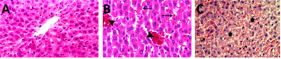
Figure 1: Histology of hepatic tissue of experimental animals (H&E; x400). A: Photomicrograph showing the normal architecture of the liver (Control group A). B: Photomicrograph of section of liver showing the effect of Malathion 27 mg/kg (Low dose group B). C: Photomicrograph showing more toxic effects of Malathion 50mg/kg (High dose group C) on liver.
Table II: Grading-wise comparison of histopathological changes in hepatic tissues of rats |
||||||
Groups |
Cellular infiltration |
Sinusoidal dilation |
Hemorrhage |
Vascular congestion |
Vacuolar degeneration |
Necrosis |
A |
0 |
0 |
0 |
0 |
0 |
0 |
B |
I |
II |
I |
II |
0 |
0 |
C |
III |
III |
III |
II |
II |
III |
Group A-Control group; Group B-low dose Malathion group; Group C-High dose Malathion group Grading score follows: none (0), mild (I), moderate (II) and severe (III)
The use of pesticides like Malathion leads to its lingering effects in the environment for a variable span. This residue of Malathion contaminates plants, crops, water as well as other food sources of humans and animals, and therefore considered an eminent chronic hazard to both18.
The current study compares the changes in the body weight, liver weight and the morphological changes in the liver of rats exposed to Malathion with unexposed rats. There was a significant reduction in the body-weight gain of Malathion-treated groups as compared with the control group, and more so in the group receiving high dose of the toxic agent. On the other hand, a significant increase in the relative weight of the liver in the treated groups was observed. These findings are consistent with Selmiet al., who also demonstrated the alterations in morphometric parameters in their studies following Malathion induction19.
These morphological variations may result from the characteristic of inhibition of acetylcholinesterase enzyme that includes the accumulation of acetylcholine resulting in stimulation of different receptors (nicotinic, cholinergic and muscarinic receptors) and neurological alterations in Malathion treated group of rats20n.
The main objective of the present study was to observe the hepatotoxic effects of different doses of Malathion. None of the rats died during the duration of the experiment. We observed moderate to severe histopathological in the mentioned doses of Malathion. Different studies also observed the toxic effects of Malathion on hepatic tissues of rats. A study conducted by Hosseini et al. observed similar hepatotoxic changes in their experimental animals with the use of Malathion21. Another study by Gupta et al. reported disorganization of the normal hepatic architecture after malathion exposure.17 While Severcan et al. observed the hepatic as well as biochemical alterations in their rats after giving Malathion 100mg/kg, 200mg/kg and 400mg/kg22. Abdel Salam et al. also demonstrated the deleterious effect of Malathion on the liver primarily causing structural and functional disruption in hepatocytes23.
The present study did not observe the hematological effects of Malathion on rat’s blood which is one of the limitations of this study. Several other studies observed that Malathion induction results in the alterations of hematological parameters in clinical cases as well as in the experimental animals. Malathion induction results in leukocytosis which is directly linked with response to the state of stress by the immune system12, 17. Findings of the present study support this possibility as histopathological analysis of our study also observed moderate-to-severe hemorrhages, infiltration of inflammatory cells, vascular congestions, and necrosis.
The histopathological findings observed in the current study included inflammatory cell infiltrations, sinusoidal and vascular congestion, hepatocyte hypertrophy, vacuolization, nuclear pyknosis, the disintegration of the hepatocyte membrane and vascular hemorrhages. With regards to such histopathological analysis, our study findings are consistent with the study by Hosseini et al, Gupta et al., Severcan et al. and Abdel Salam et al., who observed similar findings in the liver of their study animals following Malathion induced hepatotoxicity17, 21, 22.
The foremost limitation was the constraint of time and resources. Other parameters like hematological parameters, hepatic markers, and oxidative stress markers were not researched.
Based on the findings of the present study, it can be concluded that Malathion is a potent toxic pesticide and exposure to it can result in dose dependent damage to hepatic tissues.
Further studies are recommended for a more detailed evaluation of the toxic effects of Malathion on other organs as well as other parameters that may give more insight into the topic.
- Mulla SI, Ameen F, Talwar MP, Eqani SA, Bharagava RN, Saxena G, et al. Organophosphate pesticides: impact on environment, toxicity, and their degradation. Bioremediation of Industrial Waste for Environmental Safety: Springer; 2020. p. 265-90.
- Ibrahim A. Biochemical and histopathological response of Oreochromis niloticus to malathion hepatotoxicity. J Royal Sci. 2019; 1(1): 10-5.
- Ferreira RCB, Papini S, Luchini LC, Vieira E. Persistence of malathion used in dengue control on household surfaces. Arq Ins Biol. 2019; 86.
- Lasram MM, Annabi AB, El Elj N, Selmi S, Kamoun A, El-Fazaa S, et al. Metabolic disorders of acute exposure to malathion in adult Wistar rats. J Hazard Mater. 2009; 163(2-3): 1052-5. Doi: 10.1016/j.jhazmat.2008.07.059.
- Azmi M, Naqvi S, Azmi M. Pesticide residue in the blood of rural population from Gadap, Karachi and related health hazards. J Exp Zool. 2005; 8: 343-51.
- Bhalli JA, Khan Q, Haq M, Khalid A, Nasim A. Cytogenetic analysis of Pakistani individuals occupationally exposed to pesticides in a pesticide production industry. Mutagenesis. 2006; 21(2): 143-8. Doi: 10.1093/mutage/gel009.
- Walia A, Sumal K, Kumari S. Effect of chlorpyrifos and malathion on soil microbial population and enzyme activity. Acta Sci Microbiol. 2018; 1(4): 14-22. Doi: 10.31080/ASMI.2018.01.0033.
- Geng X, Shao H, Zhang Z, Ng JC, Peng C. Malathion-induced testicular toxicity is associated with spermatogenic apoptosis and alterations in testicular enzymes and hormone levels in male Wistar rats. Environ Toxicol Pharmacol. 2015; 39(2): 659-67. Doi: 10.1016/j.etap.2015.01.010.
- Alsherbiny MA, Abd-Elsalam WH, Taher E, Fares M, Torres A, Chang D, et al. Ameliorative and protective effects of ginger and its main constituents against natural, chemical and radiation-induced toxicities: a comprehensive review. Food Chem Toxicol. 2019; 123: 72-97. Doi: 10.1016/j.fct.2018.10.048.
- Abdel-Daim MM, Abushouk AI, Bungău SG, Bin-Jumah M, El-Kott AF, Shati AA, et al. Protective effects of thymoquinone and diallyl sulphide against malathion-induced toxicity in rats. Environ Sci Pollut Res Int. 2020; 27(10): 10228-35. Doi: 10.1007/s11356-019-07580-y.
- Yokota K, Fukuda M, Katafuchi R, Okamoto T. Nephrotic syndrome and acute kidney injury induced by malathion toxicity-Case Reports. BMJ Case Rep. 2017: bcr-2017-220733. Doi: 10.1136/bcr-2017-220733.
- Aboubakr HM, Elzohairy EA, Ali AA, Rashed LA, Elkady NK, Soliman AS. Therapeutic effects of N-acetylcysteine against malathion-induced hepatotoxicity. Egypt J Forensic Sci. 2019; 9(1): 34. Doi: 10.1186/s41935-019-0142-6.
- Esen M, Uysal M. Protective effects of intravenous lipid emulsion on malathion-induced hepatotoxicity. Bratisl Lek Listy. 2018; 119(6): 373-8. Doi: 10.4149/BLL_2018_069.
- Charan J, Kantharia N. How to calculate sample size in animal studies? J Pharmacol Pharmacotherap. 2013; 4(4): 303-06. Doi: 10.4103/0976-500X.119726.
- Kalender S, Uzun FG, Durak D, Demir F, Kalender Y. Malathion-induced hepatotoxicity in rats: the effects of vitamins C and E. Food Chem Toxicol. 2010; 48(2): 633-8. Doi: 10.1016/j.fct.2009.11.044.
- Elzoghby RR, Ahlam FH, Abdel-Fatah A, Farouk M. Protective role of vitamin C and green tea extract on malathion-induced hepatotoxicity and nephrotoxicity in rats. American J Pharmacol Toxicol. 2014; 9(3): 177. Doi: 10.3844/ajptsp.2014.177.188.
- Gupta VK, Siddiqi NJ, Ojha AK, Sharma B. Hepatoprotective effect of Aloe vera against cartap‐and malathion‐induced toxicity in Wistar rats. J Cell Physiol. 2019; 234(10): 18329-43. Doi:10.1002/jcp.28466
- Salih SI, Karem KK, Alhussain HAAA, Al-hindawi AA. Protective Effect of Green Tea Against Poisoning with Malathion in Adult Rats. Indian J Pub Health Res Dev. 2019; 10(2): 716-20. Doi: 10.37506/ijphrd. v10i2.7714.
- Selmi S, Rtibi K, Grami D, Sebai H, Marzouki L. Malathion, an organophosphate insecticide, provokes metabolic, histopathologic and molecular disorders in liver and kidney in prepubertal male mice. Toxicol Rep. 2018; 5: 189-95. Doi: 10.1016/j.toxrep.2017.12.021.
- Taherdehi FG, Nikravesh MR, Jalali M, Fazel A. Evaluating the protective effects of vitamin C on serum and erythrocyte cholinesterase activity of male rats exposed to malathion. Electron Physician. 2016; 8(7): 2633-8. Doi: 10.19082/2633.
- Hosseini SA, Ahmadipour A, Soltani M, Mehdipour M, Mandegary A, Karami-Mohajeri S. Malathion-increased Hepatotoxicity in Diabetic Rats. Pharma Biomed Res. 2020; 6(1): 53-60. Doi: 10.18502/pbr.v6i1.3428.
- Severcan Ç, Ekremoglu M, Sen B, Pasaoglu OT, Akyurek N, Severcan SM, et al. Acute effects of different doses of malathion on the rat liver. Clin Exp Hepatol. 2019; 5(3): 237-43. Doi: 10.5114/ceh.2019.87637.
- Abdel-Salam OM, Sleem AA, Youness ER, Morsy FA. Preventive effects of cannabis on neurotoxic and hepatotoxic activities of malathion in rat. Asian Pacific J Trop Med. 2018; 11(4): 272-9. Doi: 10.4103/1995-7645.231467.

An Official Publication of
Islamabad Medical & Dental College
Volume 10 Issue 1
Author's Contribution:
1Conception; Literature research; manuscript design and drafting; 2,3 Critical analysis and manuscript review; 4,5Data analysis; Manuscript Editing.
Article info:
Received: April 16, 2020
Accepted: December 17, 2020
Funding Source: Nil
Conflict of Interest: Nil
Address of Correspondence
Mansoor Mukhtar Qazi
Email:
dr.lumhs119@yahoo.com
Cite this article. Channar SP, Qazi N, Almani SA, Memon SG, Qazi MM, Qureshi R. Histomorphometric Alterations in Hepatic Tissue from Malathion-Induced Toxicity: An Experimental Animal Study. J Islamabad Med Dental Coll. 2021; 10(1): 56-61. Doi: 10.35787/jimdc.v10i1.534