Impact of Caffeine and Vitamin D3 on the Development of Neonate Mice Femur Growth Plate
Maimoona Khan1, Abdullah Qamar2, Ayesha Ali3, Faiza Umbreen4, Fareeha Mushtaq 5
1 Assist Prof Anatomy, Mohi-ud-Din Islamic Medical College, AJK
2, 4Assist Prof Anatomy, Army Medical College, Rawalpindi
3PG Trainee, Army Medical College, Rawalpindi
5Assist Prof Anatomy, Mohtarma Benazir Bhutto Shaheed Medical College, AJK
Background: High dosage of caffeine can influence histological changes during bone development in the newborns, while vitamin D3 has also its effect on such changes. Main objectives of the study were to determine the effects of caffeine on the histomorphology of developing femur and the role of vitamin D3 along with caffeine on the femur of BALB/c mice.
Methodology: The animal experimental study was carried out in the National Institute of Health, Islamabad in collaboration with the Anatomy Department, Army Medical College, Rawalpindi, from October 2014 till October 2015. Thirty pregnant mice, weighing 26-28g, were chosen and grouped into 3 equal sets of ten mice each. Control group G1 was fed normal diet with ad libitum access to water. Experimental group G2, along with the above diet, was provided caffeine at 10mg/100g body weight as a single dose on every second day using oral gavage for 3 weeks. Experimental group G3 was administered with caffeine at 10mg/100g body weight on every second day along with vitamin D3 0.1µg/day for 3 weeks. At completion of the study, neonate mice femurs were analysed to see the changes on the proliferative and hypertrophy zone heights of growth plate.
Results: Proliferative and hypertrophy zones of control group G1 mice mean height ±SD was measured as 540±10.99μm and 164±6.609μm, respectively, while for experimental group G2 the same height was observed as 443.5±12.258μm and 138.25±6.129μm, respectively. For experimental group G3 mice mean height ± SD of these zones was found as 474±3.839μm and 144.25±3.726μm, respectively.
Conclusion: Ingestion of caffeine modified the femur's proliferative and hypertrophy zones height of the growth plate; however, vitamin D3 dosage mitigated this consequence.
Keywords: Caffeine, Femur, Hypertrophy zone, Proliferative zone, Vitamin D3
Caffeine is one of the major components in tea, coffee, carbonated drinks, energy drinks, chocolate derived from cocoa seads1, and it is the most common psychoactive drug(legal) in the world. The role of caffeine in improving pain has been less known in the past, but now its being increasingly considered2.
Worldwide utilization of caffeine has been evaluated at 120,000 tons per year3.
At present, a great rise in soft drinks ingestion has been seen throughout the world resulting in caffeine abuse; this is especially due to massive publicity campaign and lack of appropriate understanding regarding outcomes of excessive caffeine use4. Experimental studies on young animals have reported that caffeine causes impaired bony ossification and maturation. Moreover, it impedes osteoblast differentiation, formation as well as mineralization of extracellular bony matrix5.
Previous research works have investigated that ingesting more than 500-600mg of caffeine in a single day might cause agitation, sleeplessness, anxiety, irritability, nervousness, an upset appetite, tachycardia and even muscle twitching6. In females, the half life of caffeine is approximately 5.2 to 5.4 hrs, which becomes longer during pregnancy. The consumption of 300mg of caffeine per day during gestation may potentiate the risk of premature fetal births, reduced crown rump length, micrognathia, cleft palate, limb malformations, hydrocephalus and in severe cases may even cause early death of the developing fetus7. The metabolism of caffeine is relatively slower in pregnant females, fetuses, infants and chronic hepatic illnesses8.
Vitamin D exists in two forms, specifically ergocalciferol (vitamin D2) – gotten from plants and cholecalciferol (vitamin D3) - in all other dietary sources including skin production of vitamin D9. Vitamin D performs a fundamental role in controlling as well as sustaining the mineralization of osseous tissue in all age groups and plays a central role in the regulation of mineral homeostasis and skeletal health10.
The previous experimental studies have not sufficiently focused on the histology of the developing long bones in the proliferative and hypertrophy phases. Consequently, the combined effect of caffeine and vitamin D consumption on these developing histological zones has not been investigated. Therefore, objective of this experiment was to find the outcome of caffeine consumption as well as the added effect of vitamin D3 on the growth plate of neonate femur of BALB/c mice.
The animal experimental study was conducted in the National Institute of Health (NIH), Islamabad in collaboration with the Anatomy Department, Army Medical College, Rawalpindi from October, 2014 till October, 2015 after necessary approval from ethical-committee (No. 02 / CREAM-A / 11 Feb, 2015). Convenience based non-probability sampling technique was used. Healthy pregnant mice, weighing 26-28g, total thirty (30) in number, were taken for the experiment. Non-pregnant and un-healthy female mice were not used in this research. Room environment was controlled between 20-260C. Using lottery method, pregnant mice were divided into three equal group sets of 10. Control group G1 was provided with usual laboratory food for 3 weeks. Experimental group G2 was given pure caffeine of 10mg/100g body weight, on every second day for 3 weeks, using oral gavage needle. Group G3 was administered caffeine 10mg/100g body weight on every second day, three days a week along with vitamin D3 0.1µg per day, also through oral gavage needle for 3 weeks. After 21 days of gestation period, the animals were euthanized using ether as anesthesia. When mouse became unconscious, it was placed on a clean sheet of paper on a dissecting board. The abdominal cavity was opened and gravid uterus was exposed to take foetuses out. The total number of foetuses within the uterus of all the animals were counted as 206. Two foetuses were taken randomly from each pregnant animal and right femur of newborns was dissected by separating from hip and knee joints. Fixation of femurs was done in 10% formalin and the bottles were marked appropriately. The samples were decalcified through 5% Nitric acid solution for 18-24 hrs11. Later, further processing was done in Leica TP 1020 automatic tissue processor. Infiltration and embedding was performed in paraffin wax using embedding center LEICA EG 1160. Rotary microtome was used to make 5µm thickness longitudinal bone tissue sections on warm water bath at 450C and slides were prepared. Staining using Leica auto-stainer X in hematoxylin and eosin was performed in Pathology Laboratory of AMC. Heights of proliferative and hypertrophy zones were measured through ocular micrometer (40X lens). Average of three readings in the central portion of each zone was noted down as the height of the zone12. IBM-SPSS version 20 was used for analyses using ANOVA test, while for inter-group comparison of quantitative variables, Post Hoc Tukey`s test was used. A p value ≤ 0.05 was taken as significant.
The mean heights ±SD of proliferative and hypertrophy zones for all the three groups (G1, G2 and G3) as well as statistical findings are given in Table 1. The height of both the zones was decreased in caffeine nourished G2 group in comparison to the control group G1 and experimental group G3 (Fig 1-3). In G3 experimental group, vitamin D3 showed a role in diminishing some of the effects of caffeine, through protection of the architecture of osseous tissue.
Table 1: Mean values of zones height of femur (growth plate) of new-borns |
||||
Zone |
Group G1 |
Group G2 |
Group G3 |
p-value |
Proliferative |
540.5±10.99 |
443.5±12.258 |
474±3.839 |
< .001* |
Hypertrophy |
164±6.609 |
138.25±6.129 |
144.25±3.726 |
< .001* |
*P value ≤ .05 is statistically significant
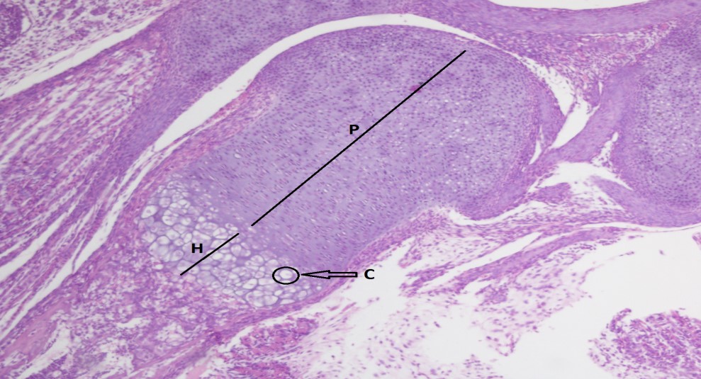
Fig 1. Control group G1, indicating proliferative zone (P), hypertrophy zone (H), chondrocyte with lacunae (C) and column of chondrocyte (Col) of femur - (40X)
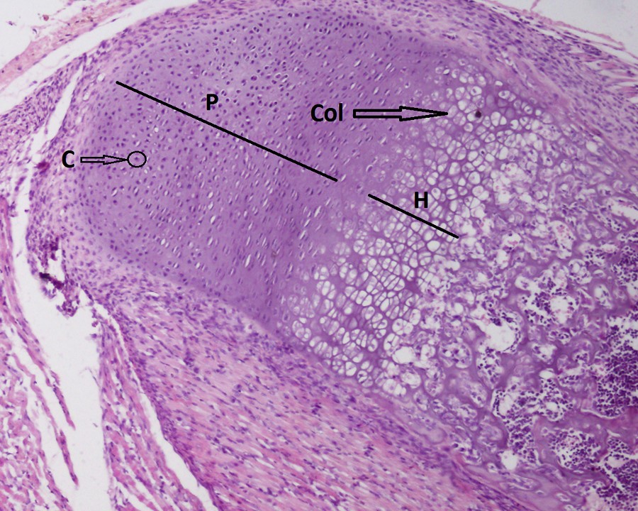
Fig 2. Experimental group G2, indicating decrease in proliferative (P) and hypertrophy (H) zones' height of femur- (40X)
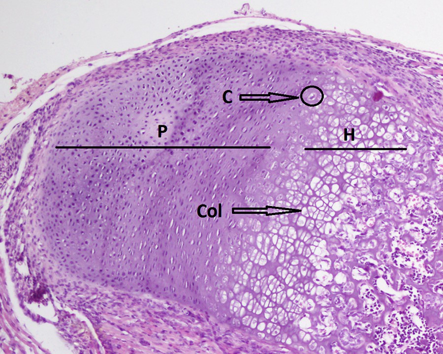
Fig 3. Experimental group G3, indicating relative increase in proliferative (P) and hypertrophy (H) zones' height of femur - (40X)
The present study showed that the height of proliferative and hypertrophy zones of the femur was appreciably reduced in caffeine nourished mice in comparison with the control group. It was determined that the caffein appeared to inhibit the action of endo-chondrocytes and reduce the thickness of epiphyseal cartilage13. Thus, in caffeine nourished mice, smaller heights of proliferative and hypertrophy zones caused lesser longitudinal growth of the femur14. Similar findings were noted in previous studies in which caffeine altered osteogenic activity, leading to impaired matrix mineralization and maturation15.
and had detrimental effects on cartilage growth, with severity of adverse effects dependent on dose16, 17. Caffeine passes into off-spring through the placenta, which causes teratogenic transformations and can reduce the formation, growth, and mass of the bones18. The mesenchymal stem cells are responsible for ossification of the whole skeleton. The caffeine that goes from the mother to the fetus during pregnancy decreases the osteogenic differentiation of mesenchymal stem cells. The reduction in the osteogenic potential of mesenchymal stem cells is implicated in the pathogenesis of osteopenia resulting from caffeine ingestion19.
In another study, significant histological changes were observed in caffeine treated rats. Thickness of epiphyseal plate (particularly proliferating and hypertrophic zones) was reduced in a group which received caffeine. The diaphysis of caffeine treated group manifested thinning out of the outer compact bone with multiple osteoporotic cavities in the bone matrix.20 The, addition of caffeine in rat chondrocytes cultures diminished cellularity, viability, and matrix synthesis activity21. These results match with the findings of current research.
Caffeine fed neonates and young rats demonstrated marked decrease in longitudinal bone growth and maturation22. Caffeine ingestion during early years resulted in harmful effects on the bony structure in the subsequent life, hence altering the bone mineral density perpetually, decreasing calcium retention and reducing height of hypertrophic zone22. Observations of the present study are also comparable. The cells of epiphyseal cartilage control the longitudinal growth of bone23. Therefore, it has been determined that high doses of caffeine can inhibit endochondral ossification in young rats24.
Experimental group (G3) in the current study also showed reduction in detrimental effect of caffeine on bones due to addition of vitamin D in the diet. The role of vitamin D is critical for normal growth and mineralization of the bony skeleton of the developing fetuses25. Vitamin D intake improved maternal calcium retention from gastrointestinal tract and enhanced offspring bone mineral density26. The current study focused on observing the development of histological zones in the new-born using conventional microscope and thus did not examine the changes in shape, size and number of the cells in the respective zones. It is, therefore, recommended that electron microscope may be used to observe the ultra structural changes in the developing bone zones in future studies.
Caffeine stimulated decrease in height of proliferative and hypertrophy zones of growth plate of new born mice's femur, possibly due to inhibition of endochondral ossification. The dosage of vitamin D3, however, alleviated this negative influence of caffeine.
- Abidi S, Gilani U, Zehra R. Extraction and analysis of caffeine from various brands of tea leaves marketed in Pakistan. J Pharmacogn Phytochem. 2020;9(1):09-10. ISSN: 2278-4136
- Baratloo A, Rouhipour A, Forouzanfar MM, Safari S, Amiri M, Negida A. The role of caffeine in pain management: a brief literature review. Anesth Pain Med. 2016;6(3). Doi: 10.5812/aapm.33193
- Alsabri SG, Mari WO, Younes S, Elsadawi MA, Oroszi TL. Kinetic and dynamic description of caffeine. J Caffeine Adenosine Res. 2018;8(1):3-9. Doi: 10.1089/caff.2017.0011
- Wassef B, Kohansieh M, Makaryus AN. Effects of energy drinks on the cardiovascular system. World J Cardiol.. 2017;9(11):796. Doi: 10.4330/wjc.v9.i11.796
- Hua R, Zou J, Ma Y, Wang X, Chen Y, Li Y, et al. Psoralidin prevents caffeine-induced damage and abnormal differentiation of bone marrow mesenchymal stem cells via the classical estrogen receptor pathway. Annals of Translational Medicine. 2021;9(15).1245. Doi: 10.21037/atm-21-31530
- Sanchis-Gomar F, Pareja-Galeano H, Cervellin G, Lippi G, Earnest CP. Energy drink overconsumption in adolescents: implications for arrhythmias and other cardiovascular events. Can J Cardiol.2015;31(5):572-5. Doi: 10.1016/j.cjca.2014.12.019
- Del-Ponte B, Santos IS, Tovo-Rodrigues L, Anselmi L, Munhoz TN, Matijasevich A. Caffeine consumption during pregnancy and ADHD at the age of 11 years: a birth cohort study. BMJ open. 2016;6(12):e012749. doi: 10.1136/bmjopen-2016-012749
- Liu J, Sui X, Lavie CJ, Hebert JR, Earnest CP, Zhang J, et al. Association of coffee consumption with all-cause and cardiovascular disease mortality. In Mayo clinic proceedings. Elsevier.2013 ;88 (10):1066-1074). Doi: 10.1016/j.mayocp.2013.06.020
- Roth DE, Abrams SA, Aloia J, Bergeron G, Bourassa MW, Brown KH, et al. Global prevalence and disease burden of vitamin D deficiency: a roadmap for action in low-and middle-income countries. Ann. N. Y. Acad. Sci. 2018 Oct;1430(1):44. Doi: 10.1111/nyas.13968
- Ryan BA, Kovacs CS. Maternal and fetal vitamin D and their roles in mineral homeostasis and fetal bone development. J Endocrinol Invest. 2021;44(4):643-59. Doi: 10.1007/s40618-020-01387-2
- H Liu, Zhu R, Liu C, Ma R, Wang L, Chen B, et al. Evaluation of decalcification techniques for rat femurs using HE and immunohistochemical staining. BioMed Res Int.2017. Doi.org/10.1155/2017/9050754
- Mizuhashi K, Ono W, Matsushita Y, Sakagami N, Takahashi A, Saunders TL, et al. Resting zone of the growth plate houses a unique class of skeletal stem cells. Nature. 2018;563(7730):254-8. Doi: 10.5061/dryad.3qq5bm7
- Choi H, Choi Y, Kim J, Bae J, Roh J. Longitudinal bone growth is impaired by direct involvement of caffeine with chondrocyte differentiation in the growth plate. J Anat. 2017;230(1):117-27. Doi:10.1111/joa.12530
- Kwak Y, Choi H, Roh J. The effects of caffeine on the long bones and testes in immature and young adult rats. Toxicol Res. 2017;33(2):157-64. Doi:10.5487/TR.2017.33.2.157
- Reis AM, Oliveira KP, De Paula IH, da Silva AP, Tarrago JF, de Melo Ocarino N, et al. Nonlinear effects of caffeine on the viability, synthesis and gene expression of chondrocytes from the offspring of rats treated during pregnancy. Acta Histochemica. 2018;120(6):505-12. Doi:10.1016/j.acthis.2018.06.001
- Guillán-Fresco M, Franco-Trepat E, Alonso-Pérez A, Jorge-Mora A, López-Fagúndez M, Pazos-Pérez A, et al. Caffeine, a risk factor for osteoarthritis and longitudinal bone growth inhibition. J. Clin. Med. 2020;9(4):1163. Doi:10.3390/jcm9041163
- Tan Y, Li J, Ni Q, Zhao Z, Magdalou J, Chen L, et al. Prenatal caffeine exposure increases adult female offspring rat’s susceptibility to osteoarthritis via low-functional programming of cartilage IGF-1 with histone acetylation. Toxicology Letters. 2018; 295:229-36. Doi: 10.1016/j.toxlet.2018.06.1221
- Wikoff D, Welsh BT, Henderson R, Brorby GP, Britt J, Myers E, et al. Systematic review of the potential adverse effects of caffeine consumption in healthy adults, pregnant women, adolescents, and children. Food Chem Toxicol. 2017; 109:585-648. Doi: 10.1016/j.fct.2017.04.002
- Reis AM, Ocarino ND, Boeloni JN, Gomes DA, Goes AM, Ferreira AD, et al. Inhibition of the osteogenic differentiation of mesenchymal stem cells derived from the offspring of rats treated with caffeine during pregnancy and lactation. Connect Tissue Res. 2016;57(2):131-42. Doi:10.3109/03008207.2015.1117075
- Farag AI, Ahmad MM, Hassanein GH. Effect of carbonated soft drinks consumption on the bone of Wistar albino rat: A histomorphometric study. J Am Sci. 2016;12(8):78-84. Doi:10.7537/marsjas120816.11.
- Raines BT, Stannard JT, Stricklin OE, Stoker AM, Choma TJ, Cook JL. Effects of Caffeine on Intervertebral Disc Cell Viability in a Whole Organ Culture Model. Global Spine Journal. 2022;12(1):61-9. Doi:10.1177/2192568220948031
- Khan M, Abdullah MS, Ambreen F, Qamar K. Effect of caffeine and vitamin d3 on proliferative and hypertrophy zones of epiphyseal cartilage of mice femur. PAFMJ. 2018;68(5):1116-20.
- Haraguchi, R., Kitazawa, R., Kohara, Y., Ikedo, A., Imai, Y. and Kitazawa, S., Recent insights into long bone development: central role of hedgehog signaling pathway in regulating growth plate. Int. J. Mol. Sci.2019; 20(23):5840. Doi:10.3390/ijms20235840
- Minematsu A, Nishii Y, Imagita H, Sakata S. Long-term intake of green tea extract causes mal-conformation of trabecular bone microarchitecture in growing rats. Calcif Tissue Int. 2018;102(3):358-67. Doi:10.1007/s00223-017-0358-0
- Miliku K, Vinkhuyzen A, Blanken LM, McGrath JJ, Eyles DW, Burne TH,et al. Maternal vitamin D concentrations during pregnancy, fetal growth patterns, and risks of adverse birth outcomes. Am. J. Clin. Nutr. 2016;103(6):1514-22. Doi:10.3945/ajcn.115.123752

An Official Publication of
Islamabad Medical & Dental College
Volume 11 Issue 1
Authors’ Contribution
1Conception; Literature research; manuscript design and drafting; 2, Critical analysis and manuscript review; 3 Data analysis; 4Manuscript Editing5.
Article info
Received: June 16, 2021
Accepted: March 28, 2022
Address of Correspondence
Maimoona Khan
Email:
maimoonakhan@hotmail.com
Cite this article. Khan M, Qamar A, Ali A, Ambreen F, Mushtaq F. Impact of Caffeine and Vitamin D3 on the Development of Neonate Mice Femur Growth Plate. J Islamabad Med Dental Coll. 2022; 11(1):3-7. DOI: 10.35787/jimdc.v11i1.728
Funding Source: Nil
Conflict of Interest: Nil