Risk Factors Analysis of Vascular Invasion in Hepatocellular Carcinoma
Belqees Yawar Faiz1, Sajida Shah1, Inamullah Shah2, Zainab Malik1, Adnan Arif1
1Department of Radiology, Shia International Hospital, Islamabad; 2Department of Surgery, Foundation University, Islamabad
Objective: HCC carries a high recurrence rate after resection and transplant. This depends on the extent of vascular invasion. Preoperative detection of vascular invasion affects the management of patient and determines the prognosis. Vascular invasion may have to be looked for in tumors that show certain risk factors. This study analyzes those risk factors.
Methodology: This observational study was carried out by retrieving data from 2017 to 2020. Hepatocellular cancers treated by administering chemotherapy or chemoembolization or those with necrosis exceeding 90% were not included in this study. After exclusion the number of patients included in the study was 78. Imaging modalities used for evaluation of the tumor were multi-slice CT scan and 3-Tesla MRI. Vascular invasion was cross-tabulated against risk factors including morphology, encapsulation of tumor, size and location of the tumor.
Results: Minimum age of patients was 22 years and maximum 75 years (mean = 56.76 ± 10.857). Fifty-three percent patients (n=41) revealed invasion in single or multiple portal branches. Large tumors (p=0.002), multifocal tumors (p=0.040), un-encapsulated tumors (p=0.001), infiltrative tumors (p=0.000) and tumors with raised serum alpha fetoprotein levels (p=0.016) were found to have exaggerated vascular invasion.
Conclusion: Multifocal and large hepatocellular carcinomas are likely to have more vascular invasion.
Keywords: Hepatocellular carcinoma; Vascular invasion; Imaging.
Hepatocellular carcinoma (HCC) enjoys a global ranking of fifth most common cancer.1 In spite of advances in medical knowledge and technology in the past two decades, surgical resection and hepatic transplant remain the treatments of choice. The disease carries a high recurrence rate after transplant and R0 surgical resection reaching up to 20% to 50%.2,3 This leads to poor prognosis and survival is reported to be 7 to 16 months after recurrence. Tumor recurrence after hepatic resection and transplant depends on many factors including viral replication, degree of portal hypertension and method of surgical resection, but the most important factor in early recurrence is preoperative vascular invasion of the tumor.4 Therefore, it is of paramount importance to preoperatively evaluate the extent of vascular invasion.
Preoperative detection of vascular invasion in patients with HCC has implications in managing these patients. It also helps in determining the prognosis of the disease in each individual patient. Vascular invasion may be readily picked by diagnostic imaging modalities in some patients. More often, however, it may have to be looked for in tumors that are more likely to cause vascular invasion. Certain biological or radiological features of the tumor may point to the possibility of presence of vascular invasion. Various studies have reported some tumor factors that are associated with increased risk of vascular invasion. These include tumor size5, multifocality6, infiltrative type of tumor7, and serum alpha fetoprotein level.6 These studies have not analyzed all the risk factors associated with vascular invasion in hepatocellular carcinoma. None of these studies was carried out in Asian countries outside China. This study was aimed at analysing risk factors for vascular invasion in HCC.
This observational study was conducted after approval from Institutional Review Board. Demographic data and values of parameters under study were obtained from hospital database system. Patients scanned for hepatocellular carcinoma (HCC) from 2017 to 2020 were included. Patients treated with chemotherapy or chemoembolization and those that had more than 90% necrosis in tumors were excluded. Study sample included 78 subjects.
Patients with HCC were scanned using triphasic multi-slice CT scan as imaging tool. Gadolinium-enhanced 3-Tesla MRI was employed in patients with equivocal CT findings. Study variables included tumor morphology, encapsulation, tumor location, tumor size, tumor enhancement and vascular invasion. Size of the tumors was recorded in centimeters, measured along the longest axis of the tumor. Location was described as central or peripheral in terms of segments involved.
Morphology included shape of the tumor as well as infiltrative pattern if any. Partially encapsulated tumors were categorized as unencapsulated. Tumors were described as homogenous or heterogenous depending on their enhancement pattern. Vascular invasion was recorded as invading main portal vein (MPV), right portal vein (RPV), left portal vein (LPV), and its posterior (RPVP) and anterior (RPVA) divisions. Data was analyzed using the software Statistical Package for Social Sciences v23. Descriptive statistics were reported in means or percentages. Cross-tabulation of invasion into vessels was done with variables that included morphology of the tumor, encapsulation of tumor, location and size of the tumor. Statistical significance of the findings was calculated using Chi-square test.
Male and female patients were 54 (69.2%) and 24 (30.8%) respectively. Mean age of the study population was 56.76 ± 10.857, ranging between 22 and 75 years. Mean size of the tumors was 7.29 ± 4.761 cms, ranging from 1 to 22 cms. Fifty-three percent patients (n=41) revealed vascular invasion in single or multiple portal vein branches. Difference in vascular invasion as observed for small and large tumors was statistically significant (p=0.002). Likewise, unifocal and multifocal tumors differed significantly in their extent of vascular invasion (p=0.040) (Table 1). Location of the tumor, peripheral or central, did not reveal any significant effect on vascular invasion (Fig 1). Invasion into multiple portal branches was found in tumors that were associated with high serum alpha fetoprotein levels and in unencapsulated tumors (Fig 2). This was statistically significant (0.016 and p=0.001 respectively). Likewise, multiple vessel invasion had a highly significant association with infiltrative tumors as compared to non-infiltrative ones (p=0.000). Enhancement pattern of tumors did not reveal a significant difference although tumors with typical enhancement were likely to be more invasive than atypical ones (p=0.557).
The principal finding of our study is a significant difference of vascular invasion with respect to the
Table I: Cross tabulation of risk factors against vascular invasion of HCC |
|||
Risk Factor |
Invasion |
p-Value |
|
Absent |
Present |
||
Tumor Size |
0.002 |
||
Large-sized tumors (>5 cms) (n=41) |
2 |
39 |
|
Small sized tumors (= or < 5 cms) (n=37) |
31 |
6 |
|
Focality |
0.040 |
||
Multifocal tumors (n=41) |
11 |
32 |
|
Unifocal tumors (n=37) |
22 |
15 |
|
Serum alpha fetoprotein |
0.016 |
||
Raised (n=21) |
7 |
14 |
|
Not Raised (n=20) |
12 |
8 |
|
Encapsulation |
0.001 |
||
Unencapsulated tumors (n=37) |
10 |
27 |
|
Encapsulated tumors (n=41) |
21 |
20 |
|
Infiltration |
0.000 |
||
Present (n=34) |
7 |
27 |
|
Absent (n=44) |
24 |
20 |
|
Enhancement pattern |
0.557 |
||
Typical (n=64) |
15 |
39 |
|
Atypical (n=14) |
6 |
8 |
|
size of the tumor, larger tumors being more invasive (p=0.002). While the mean size of the tumors came out to be 7.29 ± 4.761 cm, more than half of the patients included in this study had vascular invasion in the branches of portal vein. The difference observed in vascular invasion between small and large tumors might be attributed to a decreased proportion of functioning liver and the compromise burden faced by the liver as a result.8 This is in conformity with a 2019 multi-center study carried out in China by Xu et al, where size of hepatocellular carcinoma more than 5 cm was found to be accompanied with higher rate of vascular invasion.9
Secondly, results of our study indicate a significant difference in the vascular invasion between unifocal and multifocal tumors (p=0.040). This agrees favorably with 8th critical evaluation of American Joint Commission on Cancer.1010 Previously, literature did not support the association between multifocality and vascular invasion.11 Our study showed no significant association with respect to tumor location, i.e., peripheral versus central. We found a direct correlation of unencapsulated tumors with increased vascular invasion (p=0.001). This is in contrast to a recently reported increased lymph node metastasis associated with encapsulated HCC as part of a 2019 study by Yan Y et al that reported a preoperative nomogram to predict microvascular invasion. 12 In our study, high serum levels of AFP were associated with multiple vessel invasion (p=0.016). This is in conformity with a 2020 study by Si YQ et al that has reported a positive correlation between the two (p=0.014).13
The typical enhancement of HCC on CT scan is a pattern of arterial enhancement, greater than surrounding liver parenchyma (wash-in), and lower density or signal intensity compared to the surrounding liver (wash-out) in the venous phase.14
Our study showed that HCC with typical enhancement pattern had a stronger association with vascular invasion but the association was not significant enough statistically (0.557). The finding that vascular invasion is associated more with infiltrative types of HCC is in agreement with available literature.15 The results of our study have a bearing on predicting vascular invasion of HCC which in turn, is the most potent predictor of prognosis in the patients.4 This study has re-examined the factors associated with multiple vascular invasions in HCC as seen on CT scan. Definitive diagnosis of HCC is most often based on imaging rather than histopathology. The Liver Imaging Reporting and Data System (LI-RADS), a system endorsed by American College of Radiology,
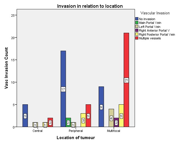
Figure 1: Comparison of vascular invasion depending on location and multifocality of HCC
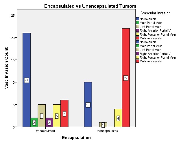
Figure 2: Comparison of vascular invasion of capsulated and unencapsulated HCC (p=0.001)
Reporting on HCC must take into account vascular invasion of the tumor in order to facilitate decision making in surgical resection. This is facilitated by identifying CT scan features that are associated with vascular invasion.
- Miller KD, Fidler-Benaoudia M, Keegan TH, Hipp HS, Jemal A, Siegel RL. Cancer statistics for adolescents and young adults, 2020. CA Cancer J Clin. 2020 Nov;70(6):443-459. https://doi.org/10.3322/caac.21637. Epub 2020 Sep 17.
- Sarici B, Isik B, Yilmaz S. Management of Recurrent HCC After Liver Transplantation. J Gastrointest Cancer. 2020 Dec;51(4):1197-1199. https://doi.org/ 10.1007/s12029-020-00498-6.
- Erstad DJ, Tanabe KK. Prognostic and Therapeutic Implications of Microvascular Invasion in Hepatocellular Carcinoma. Ann Surg Oncol. 2019 May;26(5):1474-1493. https://doi.org/ 10.1245/s10434-019-07227-9. Epub 2019 Feb 20.
- Wang W, Guo Y, Zhong J, Wang Q, Wang X, Wei H, Li J, Xiu P. The clinical significance of microvascular invasion in the surgical planning and postoperative sequential treatment in hepatocellular carcinoma. Sci Rep. 2021 Jan 28;11(1):2415. https://doi.org/ 10.1038/s41598-021-82058-x.
- Zhou L, Rui JA, Zhou WX, Wang SB, Chen SG, Qu Q. Edmondson-Steiner grade: A crucial predictor of recurrence and survival in hepatocellular carcinoma without microvascular invasion. Pathol Res Pract. 2017 Jul;213(7):824-830. https://doi.org/ 10.1016/j.prp.2017.03.002. Epub 2017 Mar 8. PMID: 28554743.
- Lei Z, Li J, Wu D, Xia Y, Wang Q, Si A, Wang K, Wan X, Lau WY, Wu M, Shen F. Nomogram for Preoperative Estimation of Microvascular Invasion Risk in Hepatitis B Virus-Related Hepatocellular Carcinoma Within the Milan Criteria. JAMA Surg. 2016 Apr;151(4):356-63. https://doi.org/10.1001/jamasurg.2015.4257. PMID: 26579636.
- He J, Shi J, Fu X, Mao L, Zhou T, Qiu Y, Zhu B. The Clinicopathologic and Prognostic Significance of Gross Classification on Solitary Hepatocellular Carcinoma After Hepatectomy. Medicine (Baltimore). 2015 Aug;94(32):e1331. https://doi.org/ 10.1097/MD.0000000000001331. PMID: 26266378; PMCID: PMC4616685.
- Ren, M et al. Liver function and energy metabolism in hepatocellular carcinoma developed in patients with hepatitis B-related cirrhosis. Medicine. 2019; 98(19): e15528-e15528. https://doi.org/ 10.1097/MD.0000000000015528.
- Xu XF, Xing H, Han J, Li ZL, Lau WY, Zhou YH, Gu WM, Wang H, Chen TH, Zeng YY, Li C, Wu MC, Shen F, Yang T. Risk Factors, Patterns, and Outcomes of Late Recurrence After Liver Resection for Hepatocellular Carcinoma: A Multicenter Study From China. JAMA Surg. 2019 Mar 1;154(3):209-217.
- Kamarajah SK, Frankel TL, Sonnenday C, Cho CS, Nathan H. Critical evaluation of the American Joint Commission on Cancer (AJCC) 8th edition staging system for patients with Hepatocellular Carcinoma (HCC): A Surveillance, Epidemiology, End Results (SEER) analysis. J Surg Oncol. 2018 Mar;117(4):644-650. https://doi.org/ 10.1002/jso.24908
- Al-Azzawi Y, Rouanet E, Hendrix RJ, Spaho L, Malik H, Devuni D, Szabo G, Barnard G. Segmental distribution of hepatocellular carcinoma correlates with microvascular invasion in liver explants undergoing transplantation. J Cancer Epidemiol. 2019 May 2;2019:8534372. https://doi.org/ 10.1155/2019/8534372. eCollection 2019.
- Yan Y, Zhou Q, Zhang M, Liu H, Lin J, Liu Q, Shi B, Wen K, Chen R, Wang J, Mao K, Xiao Z. Integrated nomograms for preoperative prediction of microvascular invasion and lymph node metastasis risk in hepatocellular carcinoma patients. Ann Surg Oncol. 2020 May;27(5):1361-1371. https://doi.org/ 10.1245/s10434-019-08071-7.
- Si YQ, Wang XQ, Fan G, Wang CY, Zheng YW, Song X, et al. Value of AFP and PIVKA-II in diagnosis of HBV-related hepatocellular carcinoma and prediction of vascular invasion and tumor differentiation. Infect Agent Cancer. 2020;15(1):70. https://doi.org/10.1186/s13027-020-00337-0.
- Ayuso C, Rimola J, Vilana R, Burrel M, Darnell A, García-Criado Á, Bianchi L, Belmonte E, Caparroz C, Barrufet M, Bruix J, Brú C. Diagnosis and staging of hepatocellular carcinoma (HCC): current guidelines. Eur J Radiol. 2018 Apr;101:72-81. Erratum in: Eur J Radiol. 2019;112:229. https://doi.org/ 10.1016/j.ejrad.2018.01.025.
- Vernuccio F, Porrello G, Cannella R, Vernuccio L, Midiri M, Giannitrapani L, et al. Benign and malignant mimickers of infiltrative hepatocellular carcinoma: tips and tricks for differential diagnosis on CT and MRI. Clin Imaging. 2021 Feb;70:33-45. https://doi.org/10.116/j.clinimag.2020.10.011.
- Tang A, Bashir MR, Corwin MT, et al. Evidence Supporting LI-RADS Major Features for CT- and MR Imaging-based Diagnosis of Hepatocellular Carcinoma: A Systematic Review. Radiology. 2018;286(1):29-48. https://doi.org/ 10.1148/radiol.2017170554.
- Song SK, Jung WY, Park SK, Chung CW, Park Y. Significantly ifferent expression levels of microRNAs associated with vascular invasion in hepatocellular carcinoma and their prognostic significance after surgical resection. PLoS One. 2019 Sep 12;14(9):e0216847. https://doi.org/ 10.1371/journal.pone.0216847.

An Official Publication of
Islamabad Medical & Dental College
Volume 13 Issue 1
Address of Correspondence
Inamullah Shah
Email:
smiubk@gmail.com
Authors’ Contribution: 1,2Conception; Literature research; manuscript design and drafting; 3,4 Critical analysis and manuscript review; 5Data analysis; Manuscript Editing.
Cite this article. Faiz BY, Shah S, Shah I, Malik Z, Arif A. Risk Factors Analysis of Vascular Invasion in Hepatocellular Carcinoma. J Islamabad Med Dental Coll. 2024; 13(1): 88-93
DOI: https://doi.org/10.35787/jimdc.v13i1.935